

 PDF
PDF ePub
ePub Citation
 Print
Print


I. Introduction
Dentin hypersensitivity is one of the most painful and least predictably treated chronic conditions in dentistry. It has been thought to be partially caused by the opening of dentinal tubules. If the exposed dentin surfaces and the number of opened dentinal tubules are increased, the patient feels more discomfort, especially during air blast, mechanical stimulation, thermal extremes or intense osmotic stimulation. In order to decrease the hypersensitivity, various attempts at blocking the dentinal tubules or changing the content of the dentinal tubules have taken place. Several medicaments and materials have been recommended to block the dentinal tubules. However, most of them have proved to be ineffective or have a short duration.
Dederich et al.1) used the Nd:YAG laser to make the dentinal walls of the root canals smooth due to melting and recrystalizing of the dentinal surface.
Harvey et al.2) demonstrated that the treated dentin with Nd:YAG laser appeared to have melted the surface because of spherical areas seen in the SEM.
Renton-Harper et al.3) demonstrated that treatment of dentin with an Nd:YAG laser showed significant effectiveness in reducing dentin hypersensitivity.
In a clinical study, Nd:YAG laser reduced dentin hypersensitivity to air stimulation by 65% and to mechanical stimulation by 72%4).
Nd:YAG laser has been reported to cause melting of dentin and closure of exposed dentinal tubules without dentin surface cracking. The sealing depth of Nd:YAG laser on human dentinal tubules was approximately 4 microns5).
Brushing dentin surfaces with dentifrice produced rough surfaces to abrasion of the enamel and the dentin at a rate of about 0.1 and 1.5 microns/min6).
The aim of the present study was to evaluate, by scanning electron microscopy, the effect of the tooth-brushing with dentifrice on Nd:YAG laser irradiated human dentin.
II. Materials and Methods
Fifty intact molar teeth stored in distilled water (4℃) were used in this study. Prior to their preparation, soft tissue and debris were removed using an ultrasonic scaler. Sound coronal dentin was exposed with high speed handpiece and diamond bur. Samples were randomly divided into 5 groups. Samples were embedded in an orthodontic resin block, and the exposed dentin surfaces were polished with 4000 grit abrasive paper.
In the control group, the exposed dentin surfaces were etched with 37% phosphoric acid (3M. Co. St. Paul, MN, U.S.A.) for 15 seconds and rinsed with distilled water for 20 seconds. Then the dry etched dentin surfaces were evenly irradiated with an Nd:YAG laser (DYL-3000EN, SAIT Co. Korea) set in 50 mJ/pulse, 10 pulses/sec for 10 seconds using a 300 µm optic fiber held 1 mm from the surface.
In the experimental groups, the etching and lasing procedures were the same as in the control group but the specimens were subsequently brushed for 15, 45, 90 and 180 minutes with an electronic toothbrush(Braun Co. U.S.A.) using a slurry made by mixing 1 gram of dentifrice (Perio toothpaste LG. Co. Korea) with 5 cc of saline under a vertical load of 120 grams on the tooth-brush. The slurry under the brush was replaced every 3 minutes.
After treatment and rinsing with saline, all specimens were stored in the saline, dried at the dry oven (80℃), gold-coated in a high resolution sputter coater (E5400, Biorad, U.S.A.) and examined in a scanning electron microscope (JEOL, JSM-840A, Tokyo, Japan).
Five sites per each specimen were observed and photographed. The number of open dentinal tubules divided by the total number of tubules per unit area were counted and the state of remaining smear layer were evaluated in a uniformly rectangular area (16×22 µm2 per each site). To isolate the group or groups that differ from the others a multiple comparison procedure (Student-Newman-Keuls Method) was used.
III. Results
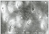
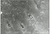
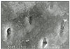
Various morphologic changes were observed on the lased and tooth-brushed dentin surfaces in the specimens.
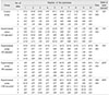
Detailed results of the number of opened dentinal tubules were shown in the Table 1.
There was no statistically significant difference between the control and the experimental groups brushed for 15 minutes or 45 minutes (P > 0.05). There was a statistically significant difference between the experimental group brushed for 45 minutes and the experimental group brushed for 90 minutes (P < 0.05).
There was a slight difference between the 90 minutes and the 180 minutes, but it was not statistically significant (P > 0.05).
IV. Discussion
Cervical abrasion is related with tooth-brushing procedures. Clinical evidence demonstrates that an excessive use of dentifrice habitually placed, undiluted, on the same area of the mouth, may produce cervical abrasion7).
Some cervical abrasive lesions may be filled with restorative material and others may be treated with desensitizing agents and materials. To treat dentin hypersensitivity, several medicaments and materials are recommended to block the dentinal tubules. The importance of occluding the dentinal tubules in certain clinical conditions is well established. The aim of therapy for sensitive teeth is to either reduce the number of open tubules or decrease their diameter8).
Brushing with a dentifrice containing calcium hydrogen phosphate as an abrasive caused most of the dentinal tubules to open6). The patency of dentinal tubules is thought to induce dentin hypersensitivity. Brushing without a dentifrice, on the other hand, resulted in occlusion of the dentinal tubules with organic pellicle-containing minerals9). Furthermore, abrasion did not occur; however, no bacterial plaque or dental calculus were retained on such surfaces. Such occluded tubules, which were distributed over the entire dentin surfaces, would likely prevent hypersensitivity. Such organic-mineral materials occluding the tubules can be derived from saliva and brushed on dentin by the brushing without using dentifrice. Kuroiwa et al. suggested that use of a non-abrasive dentifrice would prevent or reduce dentin hypersensitivity in the cervical regions9). Remizov et al. demonstrated that no abrasive effects were detected when brushing normal teeth with water. The rate of enamel and dentin abrasion caused by tooth powder or toothpaste suspensions depended on the shape of bristle cut and particularly on the toothbrush roughness10). However, many people have used dentifrice everyday without causing abrasion. They may brush for shorter times and/or use less force than patients who experience cervical abrasion.
In Kodaka's study6), automatic brushing with and without a commercial dentifrice containing calcium hydrogen phosphate as an abrasive, was performed on ground surfaces of sound enamel and dentin in human young premolar teeth, with a load of about 120 g for 10 minutes in vitro6).
Brushing with abrasive dentifrice caused rough surfaces to appear and a rate of abrasion of the enamel and the dentin of 0.1 and 1.5 microns/min, respectively. However, when brushed with only distilled water, the enamel surfaces remained intact while the dentin surfaces became smoother. Therefore, the current study adopted the use of automatic brushing with a commercial dentifrice and the pressure on the toothbrush that was same as in the Kodaka's study.
The use of the Nd:YAG laser irradiation produced a sealing of exposed tubules, however, this surface seal was removed in about 3 minutes by brushing with abrasive dentifrice6). In the current study, brushing the laser-treated surfaces for 0, 15 and 45 minutes failed to open tubules orifices (Table 1). Only after brushing for 90 min was there a significant increase in open tubules. Since many patients only brush their teeth for 1 min, once a day, the result suggests that Nd:YAG laser treatment for dentin hypersensitivity may last at least 3 months.
In Liu's study12), specimens were brushed with an electric toothbrush for 30 minutes, without any dentifrice. This is thought to be different from the usual clinical situation because most people use dentifrice during tooth-brushing. Therefore, an abrasive-containing dentifrice was used in this study for tooth-brushing and the brushing time was determined as the following. If the 3 surfaces of the 6 parts (upper and lower anterior, right upper and lower posterior and left upper and lower posterior teeth) were brushed for 3 minutes brushing, brushing time per surfaces of each part will be estimated to be 10 seconds. Therefore, accumulated brushing time per surface of 1, 3, 6 and 12 months would be about 15, 45, 90 and 180 minutes.
The use of He:Ne+Nd:YAG treatment was reported to reduce dentin sensitivity to air by 58 percent and to mechanical stimulation by 61 percent. All teeth remained vital after laser treatment, with no adverse reactions or complications4). Clinical studies showed that Nd:YAG laser can reduce dentin hypersensitivity to air by 65% and mechanical stimulation by 72% without pulpal damage during a 3-month evaluation11). According to the results of that study, a 3 month recall check and Nd:YAG laser treatment can be recommended to patients with dentin hypersensitivity.
Further clinical studies using Nd:YAG laser and other agents combined with it to intensify the efficacy of treatment of dentin hypersensitivity are indicated.
V. Conclusions
In this experiment, Nd-YAG laser irradiation of acid-etched human dentin produced occlusion of the tubules orifices. Brushing these treated surfaces 15 or 45 min did not open any tubules. After 90 and 180 mintues of toothburshing, there was statistically a significant (P<0.05) increase in the numbers of open dentinal tubules in comparison with the control, or 15 and 45 min experimental group. There was no statistically significant difference between the 90 and 180 experimental groups. Nd-YAG laser-irradiation to dentin surfaces may be effective to keep dentinal tubules closed for between 45 and 90 days.

 XML Download