

 PDF
PDF ePub
ePub Citation
 Print
Print


Abstract
C-shaped canal configuration is very difficult to treat because that clues about preoperative canal anatomy cannot be ascertained from clinical crown morphology and limited information can be derived from radiographic examination.
This study was done to get more informations about the root and canal configuration of C-shape root by 3-dimensionally reconstructing for the purpose of enhancing success rate of endodontic treatment.
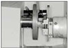
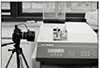
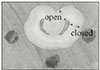
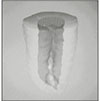
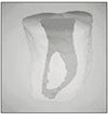
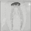
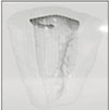
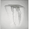
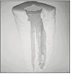
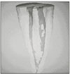
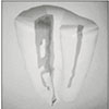
30 mandibular molars with C-shaped root were selected. Six photo images from occlusal, apical, mesial, distal, buccal, lingual directions and radiographic view were taken as preoperative ones to compare them with 3-D image. After crown reduction to the level of 1-2mm over pulpal floor was performed, teeth were stored in 5.25% sodium hypochlorite solution for the removal of pulp tissue and debris. They were cleaned under running water, allowed to bench dry and embedded in a self-curing resin. This resin block was serially ground with a microtome (Accutom-50, Struers, Denmark) and the image of each level was recorded by digital camera (FinePix S1-pro, Fuji Co., Japan). The thickness of each section was 0.25mm. Photographs of serial sections through all root canal were digitized using Adobe Photoshop 5.0 and then minimum thickness of open and closed sites were measured (open site is the surface containing occluso-apical groove : closed site is oppsite). After dizitization using 3-D Doctor (Able software Corp, USA), 3D reconstruction of the outer surface of tooth and the inner surface of pulp space was made. Canal classsification of C-shaped roots was performed from this 3-D reconstructed image.
The results were as follows:
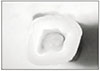
1. Most C-shape rooted teeth showed lingual groove (28/30).
2. According to Vertuccis'calssification, type I, II, III, IV, VII were observed, but also new canal types such as 2-3-2, 1-2-3-2, 2-3-2-1, 2-3-2-3 were shown.
3. There was little difference in minimum thickness on coronal and apical portions, but open site were thinner than closed site on mid portion.
Conclusively, 3D reconstruction method could make the exact configurations of C-shape root possible to be visualized and analyzed from multi-directions. Data from minimum thickness recommend cleaning and shaping be more carefully done on dangerous mid portion.
Figures and Tables


References
1. Cooke HG, Cox FL. C-shaped canal configurations in mandibular molars. J Am Dent Assoc. 1979. 99(5):836–839.

2. Manning SA. Root canal anatomy of mandibular second molars. Part II. C-shaped canals. Int Endod J. 1990. 23:40–45.
3. Weine FS. Endodontic theraphy. 1996. fifth edition. Mosby Year-Book;289.
4. Haddad GY, Nehme WB, Ounsi HF. Diagnosis, classification, and frequency of C-shaped canals in mandibular second molars in the Lebanese opulation. J Endod. 1999. 25:268–271.
5. Yang ZP, Yang SF, Lin YC, Shay JC, Chi CY. C-shaped root canals in mandibular second molars in a Chinese population. Endod Dent Traumatol. 1988. 4:160–163.
6. Weine FS, Pasiewicz RA, Rice RT. Canal Configuration of the Mandibular Seccond Molar Using a Clinically Oriented In Vitro Method. J Endod. 1988. 14:207–213.
7. Weine FS, Healey HJ, Gerstein H, Evanson L. Canal configuration in the mesiobuccal root of maxillary first molar and its endodontic significance. Oral Surg. 1969. 28:419–425.
8. Seidberg BH, Altman M, Guttuso J, Surson M. Frequency of two mesiobuccal root canals in maxillary permanent first molar. J Am Dent Assoc. 1973. 87:852–856.
9. Weine FS. The C-shaped mandibular second molar : incidence and other considerations. Members of the Arizona Endodontic Association. J Endod. 1998. 24:372–375.
10. Melton DC, Krell KV, Fuller MW. Anatomical and histological features of C-shaped canals in mandibular second molars. J Endod. 1991. 17:384–388.
11. Manning SA. Root canal anatomy of mandibular second molars. Part I. C-shaped canals. Int Endod J. 1990. 23:34–39.
12. Manning SA. Root canal anatomy of mandibular second molars. Part II. C-shaped canals. Int Endod J. 1990. 23:40–45.
13. Blaskovic-Subat V, Smojver B, Maricic B, Sutalo J. A computerized method for the evaluation of root canal morphology. Int Endod J. 1995. 28:290–296.
14. Lyroudia K, Samakovitis G, Pitas I, Lambrianidis T, Molyvdas I, Mikrogeorgis G. 3D reconstruction of two C-shape mandibular molars. J Endod. 1997. 23:101–104.
15. Lyroudia K, Mikrogeorgis G, Nikopoulos N, Samakovitis G, Molyvdas I, Pitas I. Computerized 3-D reconstruction of two "double teeth". Endod Dent Traumatol. 1997. 13:218–222.
16. Vertucci FJ. Root canal anatomy of the human permanent teeth. Oral Surg. 1984. 58:589–599.
17. Rice RT, Gilbert BO. An unusual canal configuration in a mandibular first molar. J Endod. 1987. 13:515.
18. Tamse A, Kaffe I. Radiographic survey of the prevalence of conical lower second molar. Int Endod J. 1981. 09. 14(3):188–190.
19. Bolger WL, Schindler WG. A mandibular first molar with a C-shaped root configuration. J Endod. 1988. 14:515–519.
20. Newton CW, McDonald S. A C-shaped canal configuration in a maxillary first molar. J Endod. 1984. 10:397–399.
21. Ricucci D, Pascon EA, Langeland K. Long-term follow-up on C-shaped mandibular molars. J Endod. 1996. 22:185–187.
22. Pineda F, Kuttler Y. Mesiodistal and buccolingual roentgenographic investigation of 7,275 root canals. Oral Surg. 1972. 33:101–110.
23. Carlsen O. Root complex and root canal system: a correlation analysis using one-rooted mandibular second molars. Scand J Dent Res. 1990. 98:273–285.
24. Kotoku K. Morphological studies on the roots of the Japanese mandibular second molars. Shikwa Gakuho. 1985. 85:43–64.
25. Walker RT. The root canal anatomy of mandibulars in a Southern Chinese population. J Endod. 1988. 14:325–329.
26. Jerome CE. C-shaped root canal systems: diagnosis, treatment, and restoration. Gen Dent. 1994. 42:424–427.
27. Gulabivala K, Aung TH, Alavi A, Ng YL. Root and canal morphology of Burmese mandibular molars. Int Endod J. 2001. 34:359–370.
28. Sidow SJ, West LA, Liewehr FR, Loushine RJ. Root canal morphology of human maxillary and mandibular third molar. J Endod. 2000. 26:675–678.
29. Hess W, Zeushcer E. The anatomy of the root canals of the teeth of permanent dentition. 1925. London: John Bale, Sons and Danielsson;32–34.
30. Lyroudia K, Nikolaidis N, Pitas I, Zervas P, Palakidis K. Computerized three-dimensional reconstruction: a method to study pulpal vessels and nerves. J Endod. 1993. 19:604–608.
31. Skidmore AE, Bjorndal AM. Root canal morphology of the human mandibular first molar. Oral Surg. 1971. 32:778–784.
32. Lyroudia K, Nikolaidis N, Pitas I, Palakidis K. Three computer methods to reconstruct pulpal blood vessels and nerves. J Endod. 1995. 21:501–504.
33. Lyroudia K, Palakidis K, Manthos A, Nikolaidis N, Pitas I, Foroglou C. Computerized reconstruction of TEM examined pulpal blood vessels and nerves. Endod Dent Traumatol. 1995. 11:189–195.
34. Weine FS. Endodontic theraphy on the mandibular second molar: easiest to treat of the difficult,molar teeth. Compendium. 1994. 15:1130–1628.
35. Liewehr FR, Kulild JC, Primack PD. Obturation of a C-shaped canal using an improved method of warm lateral condensation. J Endod. 1993. 19:474–477.
36. Walid N. The use of two pluggers for the obturatuion of an uncommon C-shaped canal. J Endod. 2000. 26:422–424.
37. Marshak B, Helft M, Pilo R. Factors mitigating against the use of dowels in endodontically treated teeth. Quintessence Int. 1988. 19:417–420.
38. Bolger WL, Schindler WG. A mandibular first molar with a C-shaped root configuration. J Endod. 1988. 14:515–519.
39. Bjorndal L, Carlsen O, Thuesen G, Darvann T, Kreiborg S. External and internal macromorphology in 3D-reconstructed maxillary molars using computerized X-ray microtomography. Int Endod J. 1999. 32:3–9.
40. Peters OA, Laib A, Ruegsegger P, Barbakow F. Three-dimensional analysis of root canal geometry by high-resolution computed tomography. J Dent Res. 2000. 79:1405–1409.
 XML Download