

 PDF
PDF ePub
ePub Citation
 Print
Print


Abstract
The purposes of this study were to evaluate the microtensile bond strength of one-step adhesives according to various dentin surface treatments and to observe the interface between resin(Z-100™) and dentin under SEM.
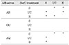
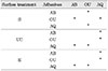
In this study forty-five non-caries extracted human molars and three adhesive systems were used; All-Bond 2(AB), One-Up Bond F(OU), AQ-Bond(AQ).; In Group 1, 2, 3, AB was used and tooth surfaces were treated by smearing(S), ultrasonic cleansing(US), etching(E) respectively. In Group 4, 5, 6, One-Up Bond F was used and tooth surfaces were also treated as the same way above. In Groups 7, 8, 9, AQ Bond was used and tooth surfaces were treated as the same way. Each specimen was prepared for microtensile bond testing, and were stored for 24hrs in 37℃ distilled water. After that, microtensile bond strength for each specimen was measured. Specimens were fabricated to examine the failure patterns of interface between resin and dentin and observed under the SEM.
The results were as follows;
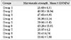
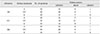
1. The results(mean±SD) of microtensile test were group 1, 25.69±4.31MPa; group 2, 40.93±10.94MPa; group 3, 47.65±8.85MPa; group 4, 36.98±9.14MPa; group 5, 39.66±8.45MPa; group 6, 43.26±13.01MPa; group 7, 25.07±4.2MPa; group 8, 30.4±4.74MPa; group 9, 33.61±7.88MPa.
2. One-Up Bond F was showed the highest value of 36.98±9.14MPa in dentin surface treatment with smearing, and there were significant differences to the other groups(p<0.05).
3. All-Bond 2 was showed the highest value of 40.93±10.94MPa in dentin surface treatment with ultrasonic cleansing, but was no significant difference to One-Up Bond F(p>0.05).
4. All-Bond 2 was showed the highest value of 47.65±8.85MPa in dentin surface treatment with etching(10%phosphoric acid), and there were significant differences to the other groups(p<0.05).
5. All-Bond 2 was showed the highest value of 47.65±8.85MPa in dentin surface treatment according to manufacture's directions, but was no significant difference to One-Up Bond F(p>0.05).
6. AQ Bond was showed the lowest microtensile bond strength with various dentin surface treatment, and the were significant differences to the other groups(p<0.05).
Figures and Tables
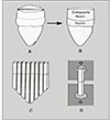
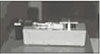
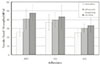
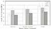
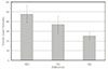
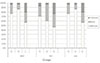
Fig. 7
All Bond 2 Smearing SEM
A:fractured dental surface, B:fractured resin surface.
1:resin, 2:dentin, 3:separated resin
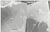
Fig. 8
All Bond 2 Ultrasonic cleaning SEM
A:fractured dental surface, B:fractured resin surface.
1:resin, 2:separated resin
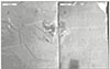
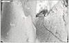
Fig. 9
All Bond 2 Etching SEM
A:fractured dental surface, B:fractured resin surface.
1:dentin, 2:bonding agent, 3:resin, 4:separated resin
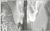
Fig. 10
One-Up Bond F Smearing SEM
A:fractured dental surface, B:fractured resin surface.
1:bonding agent, 2:resin
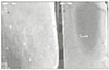
Fig. 11
One-Up Bond F Ultrasonic cleaning SEM
A:fractured dental surface, B:fractured resin surface.
1:dentin, 2:resin, 3:separated dentin
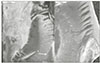
Fig. 12
One-Up Bond F Etching SEM
A:fractured dental surface, B:fractured resin surface.
1:separated dentin, 2:dentin, 3:resin, 4:dentin
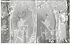
Fig. 13
AQ Bond Smearing SEM
A:fractured dental surface, B:fractured resin surface.
1:separated resin, 2:dentin, 3:resin, 4:separated dentin
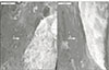
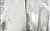
Fig. 15
AQ Bond Etching SEM
A:fractured dental surface, B:fractured resin surface.
1:dentin, 2:separated resin, 3:separated dentin, 4:resin, 5:bonding agent

References
1. Buonocore M. A simple method of increasing the adhesion of acrylic filling materials to enamel surfaces. J Dent Res. 1995. 34:849–853.

2. Pashley DH, Carvalho RM, Sano H, Nakajima M, Yoshiyama M, Shono Y, Fernandes CA, Tay F. The microtensile bond test : a review. J Adhes Dent. 1999. 1(4):299–309.
3. Schreiner RF, Chappell RP, Glaros AG, Eick JD. Microtensile testing of dentin adhesives. Dent Mater. 1998. 06. 14:194–201.
4. Chappell R, Schreiner R, Glaros A, Eixk J. Pilot study to determine sample size for micro-tensile testing. J Dent Res. 1997. 76:38. Abstr. no. 193.
5. Sano H, Sonoda H, Shono J, Takatsu T, Ciucchi B, Carvalho RM, Pashley DH. Relationship between suface area for adhesion and tensile bond strength -Evaluation of a microtensile bond test. Dent Mater. 1994. 10:236–240.
6. Sano H, Ciucchi B, Matthews WG, Russell CM, Pashley DH. Tensile properties of mineralized and demineralized human and bovine dentin. J Dent Res. 1994. 73:1205–1211.
7. Sano H, Takatsu T, Ciucchi B, Russell CM, Pashley DH. Tensile properties of resin-infiltrated demineralized human dentin. J Dent Res. 1995. 74:1093–1102.
8. Sano H, Yoshikawa T, Pereira PNR, Kanemura N, Morigami M, Tagami J, Pashley DH. Long-term durability of dentin bonds made with a self-etching primer in vivo. J Dent Res. 1999. 78:906–911.
9. Schreiner RF, Chappell RP, Glaros AG, Eick JD. Microtensile testing of dentin adhesives. Dent Mater. 1998. 14:194–202.
10. Shono Y, Terashita M, Pashley EL, Brewer PD, Pashley DH. Effects of surface area on resin-enamel tensile bond strength. Dent Mater. 1997. 13:290–296.
11. Zheng L, Pereira PN, Nakajima M, Sano H, Tagami J. Relationship between adhesive thickness and microtensile bond strength. Oper Dent. 001. Jan-Feb. 26(1):97–104.
12. Samsri S, Van Noort R. Do dentin bond strength tests serve a useful purpose? J Adhes Dent. 1999. 1:57–67.
13. Van Noort R, Noroozi S, Howard IC, Cardew G. A critique of bond strength measurements. J Dent. 1989. 17:61–67.
14. Versluis A, Tantbirojn D, Douglas WH. Why do shear bond tests pull out dentin. J Dent Res. 1997. 76:1298–1307.
15. Pashey DH, Samo H, Ciucchi B, Yoshiyama M, Carvalho RM. Adhesion testing of dentin bonding agents : A Review. Dent Mater. 1995. 11:117–125.
16. Cardoso PEC, Braga RR, Carrilho MRO. Evaluation of microtensile, shear and tensile tests determining the bond strength of three adhesive systems. Dent Mater. 1999. 14:394–398.
17. Nakabayashi N, Kojima K, Masuhara E. The promotion of adhesion by the infiltration of monomers into tooth states. J Biomed Mater Res. 1982. 16:265–273.
18. Gwinnett AJ, Matsui A. A study of enamel adhesives The Physical relationship between enamel and adhesive. Arch Oral Biol. 1967. 12:1615–1620.
19. Buonocore MG, Matsui A, Gwinnett AJ. Penetration of resin dental materials into enamel surfaces with reference to bonding. Arch Oral Biol. 1968. 13:61–70.
20. Simmelink JW, Nygaard VK, Scott DB. Penetration of resins in0to partially demineralized enamel in vivo. Arch Oral Biol. 1974. 18:435–439.
21. Gwinnet AJ, Ripa LW. Penetration of pit and fissure sealants into conditioned enamel in vivo. Arch Oral Biol. 1973. 18:435–439.
22. Wakabayashi H, Kondo Y, Suzuki K, Yatani H, Ymashita A. Effect of dissolution of collagen on adhesion to dentin. Int J Prosthodont. 1994. 7:302–306.
23. Kanca J. Wet bond. Effect of drying tome and distance. Am J Dent. 1996. 9:273–276.
24. Gwinnet AG, Tay FR, Wei SHY. Shino M, Maeda T, Suda H, Takayashi K, editors. Bridging the gap between overly dry and overwet bonding phenomenon of dentin hybridization and tubular seal. Dentin/pulp complex. 1996. Tokyo: Quintessence;359–363.
25. Suzuki M, Kato H, Wakumoto S. Vibrational analysis by Raman spectroscopy of the interface between dental adhesive resin and dentin. J Dent Res. 1991. 70:1092–1097.
26. Sano H, Shono t, Hosoda H. Microporous dentin zone beneath resin-impregnated layer. Oper Dent. 1994. 19:59–64.
27. Pashley DH, Carvalho RM. Dentin permeability and dentine adhesion. J Dent. 1997. 25:355–372.
28. Tay FR, Pashley DH. Aggressiveness of contemporary self-etching systems I: Depth of penetration beyond dentin smear layers. Dent Mater. 2001. 17:296–308.
29. Nakabayashi N, Saimi Y. Bonding to intact dentin. J Dent Res. 1996. 75:1706–1715.
30. Ferrari M, Mannocci F, Vichi A, Davidson CL. Effect of two etching times on the sealing ability of Clearfil Liner Bond 2 in Class V reatorations. Am J Dent. 1997. 10:66–70.
31. Watanable I, Nakabayashi N, Pashley DH. Bonding to ground dentin by a phenyl-P self-etching primer. J Dent Res. 1994. 73:1212–1220.
32. Hume WR. Influence of dentine on the pulpward release of eugenol or acids from restorative materials. J Oral Rehabil. 1994. 21:469–473.
33. Watanabe I, Nakabayashi N, Saimi Y. Effect of smear layer on bonding to ground dentin-relationship between grinding condition and tensile bond strength. Dent Mater J. 1994. 13:101–108.
34. Toida T, Watanabe A, Nakabayashi N. Effect of smear layer on bonding to dentin prepared with bur. Dent Mater J. 1995. 14:109–116.
35. Miyasaka K, Nakabayashi N. Combination of EDTA conditioner and Phenyl-P/HEMA self-etching primer for bonding to dentin. Dental Materials. 1999. 15:153–157.
36. Eliades G, Vougiouklakis G, Palaghias G. Heterogeneous distribution of single-bottle adhesive monomers in th resin-dentin interdiffusion zone. Dent Mater. 2001. 17:277–288.
37. Sano H, Uno S, Inous S. Sano H, Uno S, Inous S, editors. Clinical considerations of dentin adhesion. Modern trends in adhesive dentistry. 1998. Osaka: Kuraray;1–13.
38. Sano H, Takatsu T, Ciucchi B, Horner JA, Natthews WG, Pashley DH. Nanoleakage: leakage within the hybrid layer. Oper Dent. 1995. 20:18–25.
39. Sano H, Shono T, Takatsu T, Hosoda H. Microporous dentin zone beneath resin-impregnated layer. Oper Dent. 1994. 19:59–64.
40. Sano H, Yoshiyama M, Ebisu S, Burrow MF, Takatsu T, Ciucchi B, Carvalho R, Pashley DH. Comparative SEM and TEM observations of nonoleakage within the hybrid layer. Oper Dent. 1995. 20:160–170.
41. Eick JD, Robinson SJ, Chappell RP, Cobb CM, Spencer P. The dentinal surface : its influence on dentinal adhesion III. Quintessence Int. 1993. 24(8):571–574.
 XML Download