

 PDF
PDF ePub
ePub Citation
 Print
Print


Abstract
Objective
The purpose of this study was to evaluate the resin infiltration into dentin of one-bottle adhesive systems and self-etching primer bonded to Class V cavities using confocal laser scanning microscope(CLSM).
Material and Methods
Forty Class V cavities were prepared from freshly extracted caries-free human teeth. These teeth were divided into two groups based on the presence of cervical abrasion: Group I, cervical abrasion; Group II, wedge-shaped cavity preparation. Resin-dentin interfaces were produced with two one-bottle dentin bonding systems-ONE COAT BOND(OCB; Coltene®) and Syntac®Srint™(SS; VIVADENT)-, one self-etching priming system-CLEARFIL™ SE BOND(SB; KURARAY)- and one multi-step dentin bonding system-Scotchbond™Multi-Purpose(SBMP, 3M Dental Products)-as control according to manufacturers'instructions. Cavities were restored with Spectrum®(Dentsply). Specimens were immersed in saline for 24 hours and sectioned longitudinally with a low-speed diamond disc. The resin-dentin interfaces were microscopically observed using CLSM. The quality of resin-infiltrated dentin layers were evaluated by five dentists using 0-4 scale.
Results
Confocal laser scanning microscopal investigations using primer labeled with rhodamine B showed that the penetration of the primer occurred along the cavity margins.
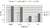
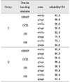
Statistical analysis using one-way ANOVA followed by Duncan's Multiple Range test revealed that the primer penetration of the group 2(wedge-shaped cavity preparation) was more effective than group 1(cervical abrasion) and that of the gingival interfaces was more effective than the occlusal interfaces. In the one-bottle dentin bonding systems, the resin penetration score of OCB was compatible to SBMP, but those of SS and self-etching priming system, SB were lower than SBMP.
Figures and Tables
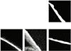
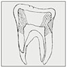 | Fig. 1Diagram of longitudinally sectioned tooth showing cavity shapes (shadowed) and schematic orientation of dentinal tubules to the class V cavity surfaces.
|
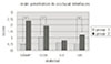 | Fig. 2Resin penetration scores in occlusal interfaces of class V restorations. Asterisk(*) means statistically significant differences(p<0.05)
|
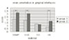 | Fig. 3Resin penetration scores in gingival interfaces of class V restorations. Asterisk(*) means statistically significant differences(p<0.05)
|
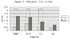 | Fig. 4Comparison of resin penetrations of occlusal and gingival interfaces in group 1(cervical abrasion). Asterisk(*) means statistically significant differences (p<0.05)
|
 | Fig. 5Comparison of resin penetrations of occlusal and gingival interfaces in group 2(V-shaped cavity preparaion). Asterisk(*) means statistically significant differences(p<0.05)
|
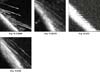
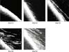 | Fig. 6.1-6.5Standard confocal laser scanning microscopic(CLSM) images at resin-dentin interfaces in occlusal interfaces of Class V cavity used for scoring the resin tag penetrations. Original magnification ×200
6.1. Score 0., 6.2. Score 1., 6.3. Score 2.,
6.4. Score 3., 6.5., Score 4.
|
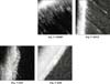
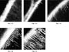 | Fig. 7.1-7.5Standard confocal laser scanning microscopic(CLSM) images at resin-dentin interfaces in gingival interfaces of Class V cavity used for scoring the resin tag penetrations. Original magnification ×200
7.1. Score 0., 7.2. Score 1., 7.3. Score 2.,
7.4. Score 3., 7.5., Score 4.
|
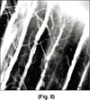 | Fig. 8Confocal laser scanning microscopic(CLSM) images at resin-dentin interfaces in gingival interfaces of Class V cavity with Scotchbond multi purpose. magnification × 800 23
|
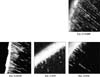
 | Fig. 9.1-9.4Confocal laser scanning microscopic(CLSM) images at resin-dentin interfaces in occlusal interfaces of cervical abrasion. Original magnification ×200
|
 | Fig. 10.1-10.4Confocal laser scanning microscopic(CLSM) images at resin-dentin interfaces in occlusal interfaces of Class V cavity. Original magnification ×200
|
 | Fig. 11.1-11.4Confocal laser scanning microscopic(CLSM) images at resin-dentin interfaces in gingival interfaces of cervical abrasion. Original magnification ×200
|
 | Fig. 12.1-12.4Confocal laser scanning microscopic(CLSM) images at resin-dentin interfaces in gingival interfaces of Class V cavity. Original magnification ×200
|
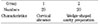
Table 2
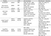
Chemical components and instructions for use of the dentin bonding systems used in this study
![]()

References
1. Pashley DH, Carvalho RM. Dentine permeability and dentine adhesion. J Dent. 1997. 25:355–372.

2. Ferrari M, Mannocci F, Cagidiaco MC, Kugel G. Short-term assessment of leakage of Class V composite restorations placed in vivo. Clin Oral Investig. 1997. 1(2):61–64.
3. Gwinett AJ, Jendersen MD. Micromorphological features of cervical erosion after acid conditioning and its relation with composite resin. J Dent Res. 1978. 57:543–549.
4. Mixon JM, Spencer P, Moore DL, Chappell RP, Adams S. Surface morphology and chemical characterization of abrasion/erosion lesions. Am J Dent. 1995. 8:5–9.
5. Heymann HO, Bayne SC. Current concepts in dentin bonding; focusing on dentinal adhesion factors. J Am Dent Assoc. 1993. 124:27–36.
6. Schupbach P, Krejci I, Lutz F. Dentin bonding: effect of tubule orientation on hybrid layer formation. Eur J Oral Sci. 1997. 105:344–352.
7. Van Meerbeek B, Braem M, Lambrechts P, Vanherle G. Morphological characterization of the interface between resin and sclerotic dentine. J Dent. 1994. 22:141–146.
8. Tay FR, Gwinnett AJ, Wei SH. Micromorphological spectrum from overdrying to overwetting acid conditioned dentin in water-free, acetone-based, single-bottle primer/adhesives. Dent Mater. 1996. 236–244.
9. Watson TF, De Wilmot DM. A confocal microscopic evaluation of the interface between Syntac adhesive and tooth tissue. J Dent. 1992. 20:302–310.
10. Duke ES, Lindemuth J. Variability of clinical dentin sustrates. Am J Dent. 1991. 4:241–246.
11. Chappell RP, Cobb CM, Spencer P, Eick JD. Dentinal tubule anastomosis: a potential factor in adhesive bonding? J Prosthet Dent. 1994. 72(2):183–188.
12. Ferrari M, Cagidiaco CM, Mason PN. Morphologic aspects of the resin-dentin interdiffusion zone with five different dentin adhesive systems tested in vivo. J Prosthet Dent. 1994. 4. 71(4):404–408.
13. Duke ES. Clinical studies of adhesive systems. Oper Dent. 1992. Suppl 5. 103–110.
14. Yoshiyama M, Carvalho R, Sano H, Horner JA, Brewer PD, Pashley DH. Regional bond strengths of resins to human root dentine. J Dent. 1996. 435–442.
15. Hannig M, Reinhardt KJ, Bott B. Self-etching primer vs phosphoric acid:An alternative concept for composite-to-enamel bonding. Oper Dent. 1999. 172–180.
16. Hannig M, Reinhardt KJ, Bott B. Self-etching primer vs phosphoric acid: An alternative concept for composite to enamel bonding. Oper Dent. 1999. 24:172–180.
17. Yoshiyama M, Sano H, Ebisu S, Tagami J, Ciucchi B, Carvalho RM, Johnson MH, Pashley DH. Regional strengths of bonding agents to cervical sclerotic root dentin. J Dent Res. 1996. 75:1404–1413.
18. Watson TF. Application of confocal scanning optical microscopy to dentistry. Br Dent J. 1991. 9:287–291.
19. Minsky M. Microscopy apparatus. 1957. Nov. 7. United States Patent Office Filed.
20. Yoshiyama M, Carvalho R, Sano H, Horner JA, Brewer PD, Pashley DH. Interfacial morphology and strength of bonds made to superficial versus deep dentin. Am J Dent. 1995. 297–302.
21. Gwinnett AJ. Quantitative contribution of resin infiltration/hybridization to dentin bonding. Am J Dent. 1993. 6:7–9.
22. Titley K, Chernecky R, Chan A, Smith D. The compositon and ultrastructure of resin tags in etched dentin. Am J Dent. 1995. 8:224–230.
23. Pashley DH, et al. Permeability of dentin to adhesive agents. Quintessence Int. 1993. 24:618–631.
24. Prati C, Chersoni S, Moniorgi R, Pashley DH. Resin-infiltrated dentin layer formation of new bonding systems. Oper Dent. 1998. 23:185–194.
 XML Download