

 PDF
PDF ePub
ePub Citation
 Print
Print


I. Introduction
Dental pulp and periradicular tissues react to bacterial infections by recruiting a variety of immunocompetent cells to the dental pulp and periradicular tissues. Pulp studies have shown the presence of immnocompetent cells and cells that recognize foreign antigens1). As a results of the interaction of microorganisms and their by-products, various mediators of inflammation, such as neuropeptides, vasoactive amines, kinins, complement component, and arachidonic acid metabolites, are released2).
It was shown that cytokines play important roles and regulate the intensity and duration of the immune response against potentially pathogenic agents. The occurrence of interleukin(IL)-1 and IL-1 producing cells has been demonstrated in human inflamed pulps3). The roles of IL-2 and IL-6 have also been studied in healthy and inflamed dental pulps4,5).
In one study on periodontal disease, the amount of IL-6 and IL-10 were significantly higher in the inflamed gingival tissues than in the peripheral blood from the healthy subjects6). IL-6 was detected in human pulps, periapical lesions5) and odontogenic cysts7). And it has been shown that the production of IL-6 was stimulated by Prevotella intermedia lipop-olysaccharide8).
Interleukin-6 is produced by mononuclear phagocytes, vascular endothelial cells, fibroblasts, and other cells in response to IL-1 and TNF9). One of the best described actions of IL-6 is on B lymphocytes. IL-6 serves as a growth factor for activated B lymphocytes late in the sequence of B cell differentiation. Activated B lymphocytes differentiate into plasma cells and produce antibody against antigens or infective microorganisms. IL-6 may serve as a cofactor of T lymphocytes and thymocytes activation. Activated T lymphocytes can produce cytokines including IL-10 that mediate inflammatory reactions. IL-6 also acts as a cofactor with other cytokines for the growth of early bone marrow hematopoietic stem cells.9) In previous studies of pulpal and periapical pathology, IL-6 is known to be one of pro-inflammatory cytokines10,11).
One of the major activities of IL-10 is to inhibit cytokine ( i.e.. TNFα, IL-1, chemokine, and IL-12) production by macrophages. It inhibits the production of IFN-γ which contribute to pathologic bone resorption in periapical lesion13). And It also suppresses the production of IL-6 by T-lymphocytes14). The effect of these actions is to inhibit T cell-mediated immune response. In addition to its inhibitory effects on macrophages, IL-10 has stimulatory actions on B lymphocytes.
Kakehashi et al. have shown that pathogenesis of pulpal and periapical lesion is closely related to microorganisms15). In their study, dental pulps of conventional and germ-free rats were exposed to their own flora. Pulpal and periradicular lesions developed in conventional rats but failed to develop in germ-free rats.
Bae et al. have shown that the most commonly isolated bacteria from infections of endodontic origin are Prevotella nigrescens16).
The purpose of this study was to determine IL-6 and IL-10 in rat pulpal inflammation experimentally induced by specific gram-negative bacteria (Prevotella nigrescens) and investigate their roles in the progress of pulpal inflammation.
II. Materials and Methods
1. Experimental induction of Pulpal inflammation
To induce pulpal inflammation experimentally on mandibular incisors of rats, we used Prevotella nigrescens(ATCC 33563) cultured on columbia agar plates supplemented with hemin and menadione at 37℃, 85% N2, 10% H2, and 5% CO2 anaerobic cha-mber.
Thirty adult male wistar rats, each weighing 250~300g were anesthetized intraperitoneally with 60 mg/kg of ketalar (Ketamine hydrochloride, Yuhan Co. Seoul). Sixty teeth were used in this experiment. The tips of lower incisors were cut off at the level of gingival margin to exclude the possibility of occlusal stress affecting the dental pulps. A hole was drilled in dentin with a round bur at 35,000 rpm without coolant to open pulp chambers. Coronal portions of the root canals were enlarged with K-files ranging from #15 to #25.
In Prevotella nigrescens groups, tiny sterile cotton pellet soaked with a bacterial colony on the agar plate was inserted into the prepared space in contact with pulp tissues. The cavities were sealed with temporary filling material (Caviton. G.C). In control groups, sterile cotton pellet without bacteria was inserted into the prepared space in contact with pulp tissues.
1, 2 and 5 days after pulpal irritation, mandibular incisors were extracted. Extracted teeth were stored in -70℃ liquid nitrogen tank.
2. Preparation of tissue samples and Protein assay
The extracted teeth were removed from the liquid nitrogen tank, and allowed to thaw for 10 minutes. After the teeth were cracked open, pulp tissues were carefully removed from the teeth. Half of the pulp tissue from one tooth was used for detection of IL-6 and the other half was used for IL-10. Pulp tissues were homogenized in buffer(0.1M potassium chloride, 0.02M TRIS; pH=7.6) in glass homogenizer and centrifuged(2000RPM, 4℃, 10min). Supernatants were collected.
The concentrations of protein in tissue sample were measured by protein assay kit.
The concentrations were measured in µg/ml tissue sample(BCA protein assay kit, Pierce, USA).
3. ELISA
The concentrations of IL-6 and IL-10 were measured using ELISA kits (Amersham Pharmacia Biotech Co. Japan). The concentrations were measured in pg/ml tissue sample.
4. Statistical Analysis
Results from ELISA were analyzed using Mann Whitney rank sum test and presented as P values. Values less than .05 were considered to be significant.
5. Histological Study
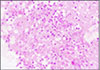
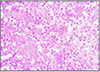
1, 2 and 5 days after pulpal irritation, rat mandibular incisors were extracted and hard tissues were decalcified with 5% nitric acid. The pulp tissues and surrounding hard tissues were longitudinally sectioned and stained by Hematoxylin and Eosin. The specimens were examined under light-microscope(×400).
III. Results
1. ELISA
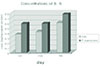
The mean concentrations of the cytokines were illustrated in table 1. Concentrations of IL-6 ranged from 0.585 to 0.778pg/µg protein in Prevotella nigrescens group and from 0.368 to 0.605pg/µg protein in the control group. Fig. 1 illustrates the concentrations of IL-6 in Prevotella nigrescens groups and control groups.
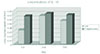
Concentrations of IL-10 ranged from 0.066 to 0.072 pg/µg protein in Prevotella nigrescens groups and from 0.033 to 0.067pg/µg protein in the control groups.
The concentrations of IL-6 and IL-10 in Prevotella nigrescens groups were higher than those in control groups on the 1st(P<0.05), 2nd and 5th day of pulpal irritation. Fig. 2 illustrates the concentrations of IL-10 in Prevotella nigrescens groups and control groups.
The concentrations of interleukin-6 in all samples were higher than those of interleukin-10.
To examine the role of IL-6 and IL-10 in the development development of pulpal inflammation, IL-10 to IL-6 ratios were calculated on the 1st, 2nd day of pulpal irritation.
IL-10 to IL-6 ratios are shown in table 2.
IL-10 to IL-6 ratios were higher on the 2nd day compared to the 1st day in the control groups (P<0.05) and Prevotella nigrescens groups.
2. Histological study
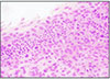
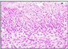
The histological study showed signs of chronic inflammatory tissue. The dental pulp tissues irritated by Prevotella nigrescens showed intense infiltration of polymorphonuclear leukocytes 2 days after pulpal irritation in Prevotella nigrescens group (Fig. 4) and control group (Fig. 3). Pulpal inflammation decreased on the 5th day of pulpal irritation in the Prevotella nigrescens group and Control group (Fig. 5, 6).
IV. Discussion
Pulp tissues in the control groups, irritated by thermal and mechanical stimulus but not exposed to bacteria, were also experimentally inflamed. We inoculated Prevotella nigrescens to examine the change of IL-6 and IL-10 production in response to the invading bacteria. The concentrations of IL-6 and IL-10 were higher in Prevotella nigrescens group than those in the control groups on the 1st, 2nd and 5th day of pulpal irritation. These findings may suggest that IL-6 and IL-10 might be involved in developing pulpal inflammation stimulated by specific bacteria. Matsushima et al demonstrated that Gram-negative bacteria, such as L. casei, from carious lesions, might be involved in developing pulpitis through the stimulation of IL-6 production17).
The results of this study were comparable to the study of Barkhordar's5), which examined the level of interleukin-6 in inflamed human dental pulps and periapical lesion. In their study, the inflamed pulpal tissues exhibited significantly higher levels of IL-6 (mean = 36±3.9pg/mg protein) compared to healthy pulp. (mean = 0.01±0.02 pg/mg protein).
In the study of Nakanishi18), differences between normal and inflamed pulp were found in the levels of IL-6 but the difference were not statistically significant.
One investigator19), examined the production of various cytokines including IL-6 and IL-10 in murine periapical inflammation. The production of both cytokines increased beginning on day 7 and increased to day 14. This results indicate that a cytokine network is activated in response to bacterial irritation and IL-6 and IL-10 played a role in the progress of periapical pathogenesis.
In this experiment, pulpal inflammation is induced instead of periapical inflammation. Histologic study demonstrated that pulpal inflammation is present on 2nd day and decreased on 5th day.
Another study20) have shown that expression of IL-6 mRNA was significantly higher in diseased periodontal tissues compared to healthy contols. They also have shown the correlation between extent of tissue damage and bone destruction.
These results were comparable to our study and support the findings that IL-6 and IL-10 were produced and released to have a role in the process of pulpal inflammation.
There are a few studies on the role of IL-6 and IL-10 in pulpal inflammation.
But the the role of IL-6 and IL-10 in the development of pulpal and periapical pathogenesis were not clearly identified.
A study of Opal SM et al., have shown that IL-6 is one of anti-inflammatory cytokine21). On the contrary, in the study of Panichi et al.10), IL-6 was known to be one of pro-inflammatory cytokines. IL-10 is known to be anti-inflammatory cytokine that suppresses the production of IL-6.
In one study it was suggested that IL-6 produced by dental pulp cells is involved in the metabolism of extracellular matrix and the destruction of dental pulp tissue17). Ishimi et al have shown that IL-6 induces bone resorption both alone and in concert with other bone-resorbing agents22). Although Lowik et al have shown that IL-6 may be a mediator in PTH-stimulated osteoclastic bone resorption23).
In this study, IL-10 to IL-6 ratio on the 2nd day were higher than that on the 1st day. At the early stage of pulpal inflammation, IL-6 was produced and it stimulated B lymphocyte to produce antibody and activate T lymphocyte to produce their own cytokines including IL-10. And in the advanced stage of pulpal inflammation, IL-10 was produced and inhibited the production of cytokines, including IL-1 and IL-613).
These results suggest that IL-6 have a role in earlier stage than IL-10 in the process of pulpal inflammation. These results may help to understand the complex regulation of T-lymphocyte mediated cytokine production by IL-10.
Further studies are necessary to elucidate the roles of IL-6 and IL-10 in developing irreversible inflammation in the dental pulp.
V. Conclusion
According to this study, we could summarize as follows:
The concentrations of interleukin-6 in Prevotella nigrescens groups were higher than those in the control groups on the 1st (P<0.05), 2nd and 5th day of pulpal irritation.
The concentrations of interleukin-10 in Prevotella nigrescens groups were higher than those in the control groups on the 1st (P<0.05), 2nd and 5th day of pulpal irritation.
IL-10 to IL-6 ratio (IL-10/IL-6) were higher on the 2nd day compared to 1st day in the control groups (P<0.05) and Prevotella nigrescens groups.
The concentrations of IL-6 were higher than IL-10 in all experimental groups and control groups.
These results suggest that Prevotella nigrescens may have a role in the progress of pulpal inflammation by stimulating the production of IL-6 and IL-10 and IL-6 may have a role in earlier stage than IL-10 in the development of pulpal inflammation.


 XML Download