

 PDF
PDF ePub
ePub Citation
 Print
Print


Abstract
One of the latest concepts in bonding are "total etch", in which both enamel and dentin are etched with an acid to remove the smear layers, and "wet dentin" in which the dentin is not dry but left moist before application of the bonding primer. Ideally, the application of a bonding agent to tooth structure should be insensitive to minor contamination from oral fluids. Clinically, contaminations such as saliva, gingival fluid, blood and handpiece lubricant are often encountered by dentists during cavity preparation.
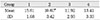
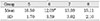
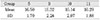
The aim of this study was to evaluate the effect of contamination by hemostatic agents on shear bond strength of compomer restorations. One hundred and ten extracted human maxillary and mandibular molar teeth were collected. The teeth were removed soft tissue remnant and debris and stored in physiologic solution until they were used. Small flat area on dentin of the buccal surface were wet ground serially with 400, 800 and 1200 abrasive papers on automatic polishing machine. The teeth were randomly divided into 11 groups. Each group was conditioned as follows:
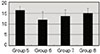
Group 1: Dentin surface was not etched and not contaminated by hemostatic agents.
Group 2: Dentin surface was not etched but was contaminated by Astringedent®(Ultradent product Inc., Utah, U.S.A.).
Group 3: Dentin surface was not etched but was contaminated by Bosmin®(Jeil Pharm, Korea.).
Group 4: Dentin surface was not etched but was contaminated by Epri-dent®(Epr Industries, NJ, U.S.A.).
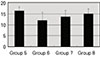
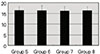
Group 5: Dentin surface was etched and not contaminated by hemostatic agents.
Group 6: Dentin surface was etched and contaminated by Astringedent®.
Group 7: Dentin surface was etched and contaminated by Bosmin®.
Group 8: Dentin surface was etched and contaminated by Epri-dent®.
Group 9: Dentin surface was contaminated by Astringedent®. The contaminated surface was rinsed by water and dried by compressed air.
Group 10: Dentin surface was contaminated by Bosmin®. The contaminated surface was rinsed by water and dried by compressed air.
Group 11: Dentin surface was contaminated by Epri-dent®. The contaminated surface was rinsed by water and dried by compressed air.
After surface conditioning, F2000® was applicated on the conditoned dentin surface. The teeth were thermocycled in distilled water at 5℃ and 55℃ for 1,000 cycles. The samples were placed on the binder with the bonded compomer-dentin interface parallel to the knife-edge shearing rod of the Universal Testing Machine(Zwick Z020, Zwick Co., Germany) running at a cross head speed of 1.0 mm/min.
Group 2 showed significant decrease in shear bond strength compared with group 1 and group 6 showed significant decrease in shear bond strength compared with group 5.
There were no significant differences in shear bond strength between group 5 and group 9, 10 and 11.
Figures and Tables
References
1. McLean JW, Nicholson JW, Wilson AD. Proposed nomenclature for glass-ionomer dental cements and realted material. Quintessence Int. 1994. 25:587–589.
2. Christensen GJ. Compomers vs resin-reinforced glass ionomers. J Am Dent Assoc. 1997. 128:479–480.

3. Buonocore MG. A simple method of increasing the adhession of acrylic filling materials to enamel surface. J Dent Res. 1955. 34:849–853.
4. Swift EJ, Perdigao J, Heymann HO. Bonding to enaeml and dentin: A brief hsitory and state of the art. Quintessence Int. 1995. 26:95–110.
5. Barkmeier WW, Los SA, Triolo PT. Bond strength and SEM evaluation of Clearfil Liner Bond 2. Am J Dent. 1997. 10:141–146.
6. Gordan VV, Vargas MA, Cobb DS, Denehy GE. Evaluation of adhesive systems using acidic primers. Am J Dent. 1997. 10:219–223.
7. Vargas MA. Interfacial ultrastructure of a self-etching primer/adhesive[abstract 95 0]. J Dent Res. 1999. 78:224.
8. Kaneshima T, Yatani H, Kasai T, Watanabe EK, Yamashita A. The influence of blood contamination on bond strengths between dentin and adhesive resin cement. Oper Dent. 2000. 25:195–201.
9. Powers JM, Finger WJ, Xie J. Bonding of composite resin to contaminated human enamel and dentin. J Prosthodont. 1995. 4:28–32.
10. Xie J, Powers JM, McGuckin RS. In vitro bond strength of two adhesives to enamel and dentin under normal and contaminated conditions. Dent Mater. 1993. 9:295–299.
11. Waston TF. A confocal optical microscope study of the morphology of the tooth/restoration using Scotchbond 2 dentin adhesive. J Dent Res. 1989. 68:1124–1131.
12. Kanca J. Resin bonding to wet substrate. I. Bonding to dentin. Quintessence Int. 1992. 23:39–41.
13. Castro AK, Hara AT, Pimenta LA. Influence of collagen removal on shear bond strength of one-bottle adhesive systems in dentin. J Adhes Dent. 2000. 2:271–277.
14. Perdiago J, Lopes M, Geraldeli S, Garcia-Godoy F. Effect of a sodium hypochlorite gel on dentin bonding. Dent Mater. 2000. 16:311–323.
15. Schwartz RS, Summitt JB, Robbins JW. Fundamental of Operative dentistry: a contemporary approach Chicage. 1996. Quintessence Pub Co;141–157.
16. Masashi M, Ko H, Hideo O, Moore BK. Influence of light intensity on shear bond strenth to dentin. Am J Dent. 1995. 8:245–248.
17. Hinoura K, Miyazaki , Onose H. Effect of irradiation time to light-cured resin composite on dentin bond strength. Am J Dent. 1991. 4:273–276.
18. Pashley EL, Tao L, Mackert JR, et al. Comparison of in vivo vs in vitro bonding of composite resin to the dentine of canine teeth. J Dent Res. 1988. 67:467–470.
19. Bruno T, Harald O, Edward J. Shear bond strengths of one-bottle adhesives to oil-contaminated enamel. J Esthet Dent. 2000. 12:139–145.
20. Ibrahim H, Franklin G. Saliva contamination and bond strength of single-bottle adhesives to enamel and dentin. Am J Dent. 1997. 10:83–87.
21. Douglas WH, Reeh ES, Raj PA, et al. A major boundary lubricant of haman saliva. Biochem Biophys Res Commun. 1991. 180:91–97.
22. Nakabayashi N, Ashizawa M, Nakamura M. Identification of a resin dentin hybrid layer in vital human dentin created in vivo: Durable bonding to vital dentin. Quintessence Int. 1992. 23:135–141.
23. Gwinnett AJ. Moist versus dry dentin: Its effect on shear bond strength. Am J Dent. 1992. 5:127–129.
24. Yamamoto T. The effect of contamination on the adhesion of composite resin to etched enamel surface. Jpn J Consev Dent. 1981. 24:93–114.
25. Tomas J, Karl J. Some effects of water on dentin bonding. Dent Mater. 1995. 11:132–136.
26. Ali I, Carel L. Bonding efficiency and interfacial morphology of one-bottle adhesives to contaminated dentin surfaces. Am J Dent. 1998. 11:281–285.
27. Woody RD, Miller A, Staffanou RS. Review of the pH of hemostatic agents used in tissue displacement. J Prosthet Dent. 1993. 70:191–192.
28. Martin F, Carla C, William M. Smear layer instability caused by hemostatic agents. J Prosthet Dent. 1996. 76:477–482.
29. Watanabe I, Nakabayashi N, Pashley DH. Bonding to ground dentin by a Phenyl-P self-etching primer. J Dent Res. 1994. 73:1212–1220.
30. Kim MS, On YS, Lee KW, Shon HH. Morphologic change of dentin surface according to the difference in concentration and application time of phosphoric acid. J Korean Acad Conserv Dent. 1998. 23:141–155.
 XML Download