

 PDF
PDF ePub
ePub Citation
 Print
Print


Abstract
Leiomyoma is a benign solitary tumor which is originated from a smooth muscle cell and grows slowly. It is most commonly found in the uterus and can develop anywhere that smooth muscle is present, including esophagus, lower extremity, stroma of GI tract, and pleura. However, the occurrence of leiomyoma in the hand is so uncommon. We present one case of solid type leiomyoma found in the right 4th finger of a young woman.
References
1. Butler ED, Hamill JP, Seipel RS, De LORIMIER AA. Tumors of the hand. A ten-year survey and report of 437 cases. Am J Surg. 1960; 100:293–302.
2. Enzinger FM, Weiss SW. Benign tumors of smooth muscle. Weiss SW, Goldblum JR, editors. Soft tissue tumors. 4th ed.St.Louis: CV Mosby;1995. p. 467–90.
3. Hachisuga T, Hashimoto H, Enjoji M. Angioleiomyoma. A clinicopathologic reappraisal of 562 cases. Cancer. 1984; 54:126–30.

4. HURWITZ A. Leiomyoma of the esophagus; report of a case. Surgery. 1949; 25:304–6.
5. Klopp EJ, Crawford BL. Leiomyoma of the small intestine. Ann Surg. 1935; 101:726–33.
6. Williams RB Jr, Daniel RA Jr. Leiomyoma of the lung. J Thorac Surg. 1950; 19:806–10.
7. Boutayeb F, El Ibrahimi A, Chraibi F, Znati K. Leiomyoma in an index finger: report of case and review of literature. Hand (N Y). 2008; 3:210–1.
8. Hwang JW, Ahn JM, Kang HS, Suh JS, Kim SM, Seo JW. Vascular leiomyoma of an extremity: MR imaging-pathology correlation. AJR Am J Roentgenol. 1998; 171:981–5.
9. Kulkarni AR, Haase SC, Chung KC. Leiomyoma of the hand. Hand (N Y). 2009; 4:145–9.
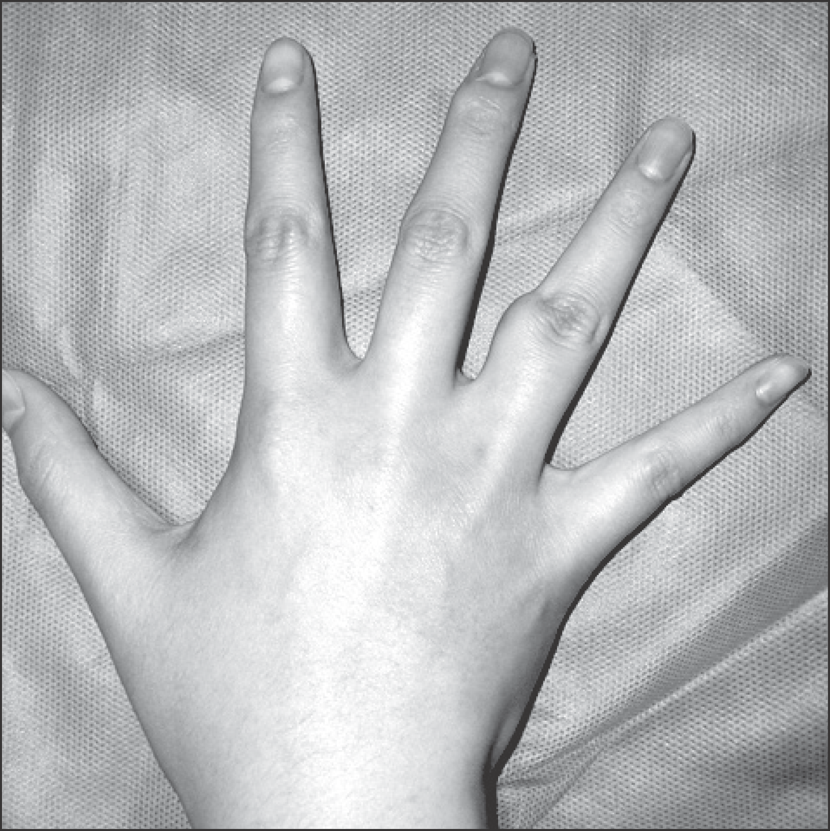
Figure 2.
Radiograph shows a round soft tissue lesion on right 4th proximal phalanx, but neither bony erosion nor calcification was observed.
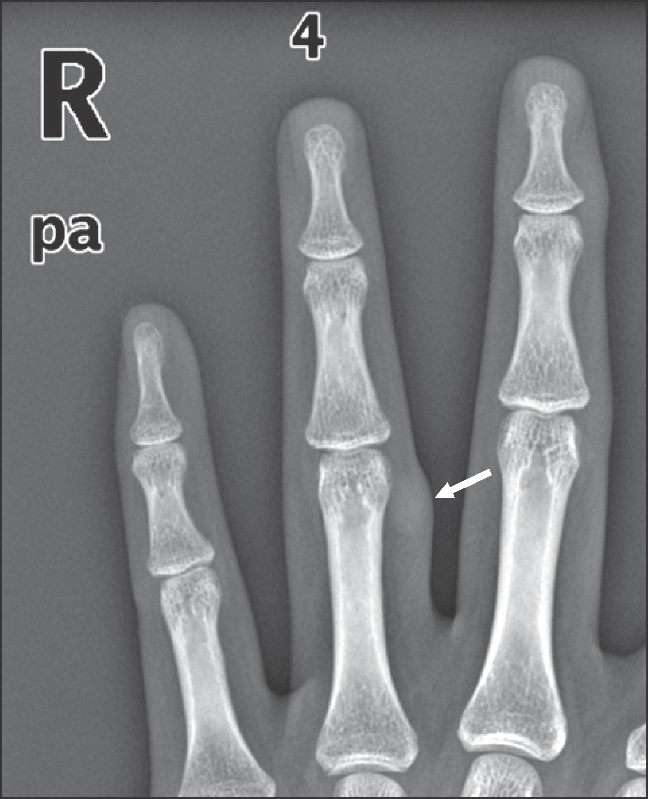
Figure 3.
Ultrasonograph shows an ovoid-shaped cystic lesion in right 4th finger proximal phalanx, which measures 0.7×0.4×0.7 cm. (A) Axial view, (B) Saggital view, (C) Doppler: No vascular flow was observed in the lesion.
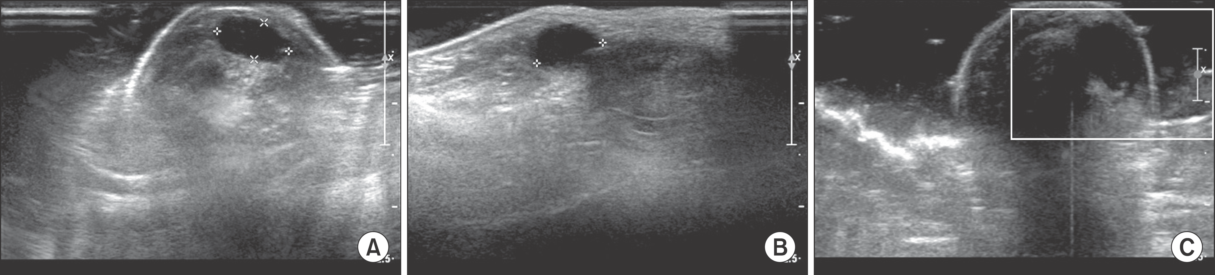
Figure 4.
Photographs show a yellow-brownish soft tissue mass. (A) The mass was dissected meticulously. (B) The mass removed was about 1×1×1 cm in size.
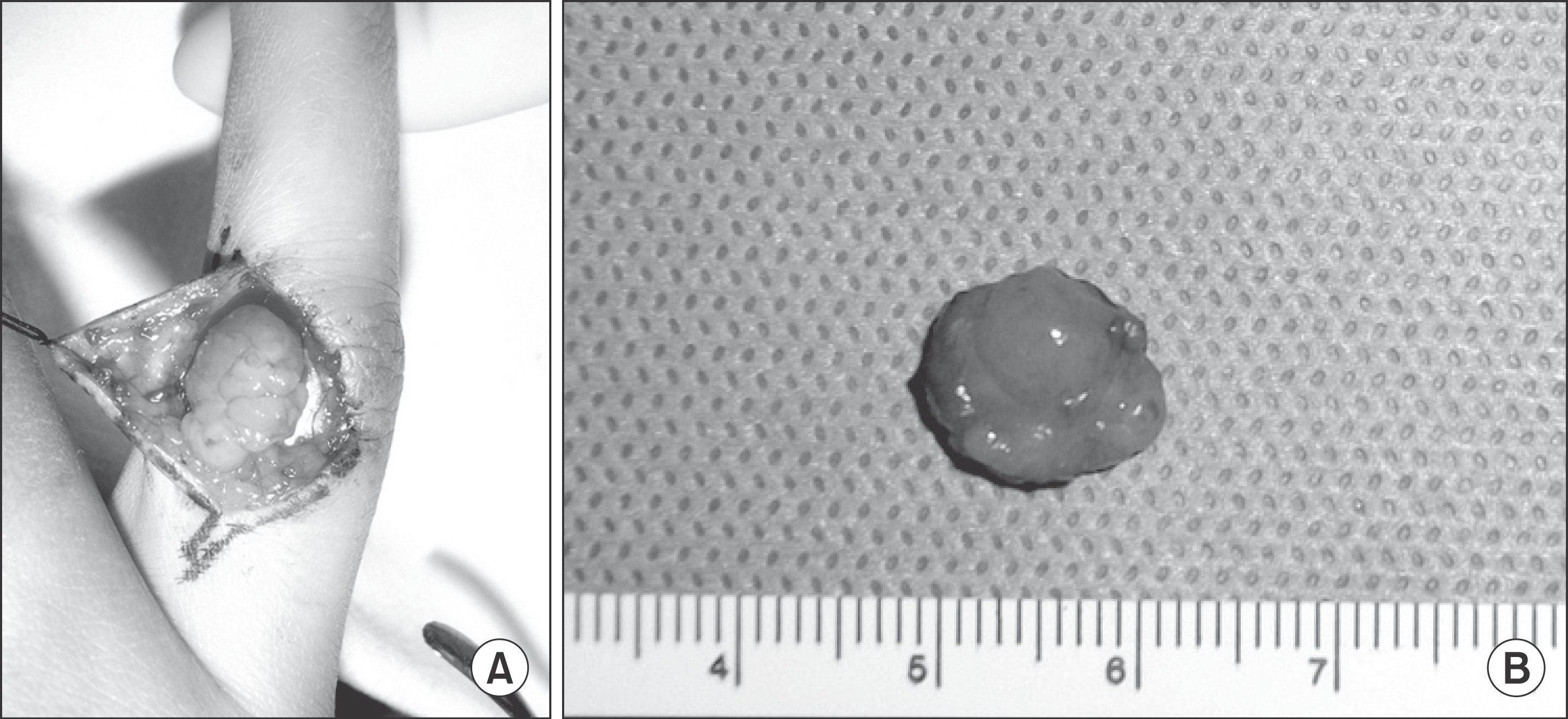
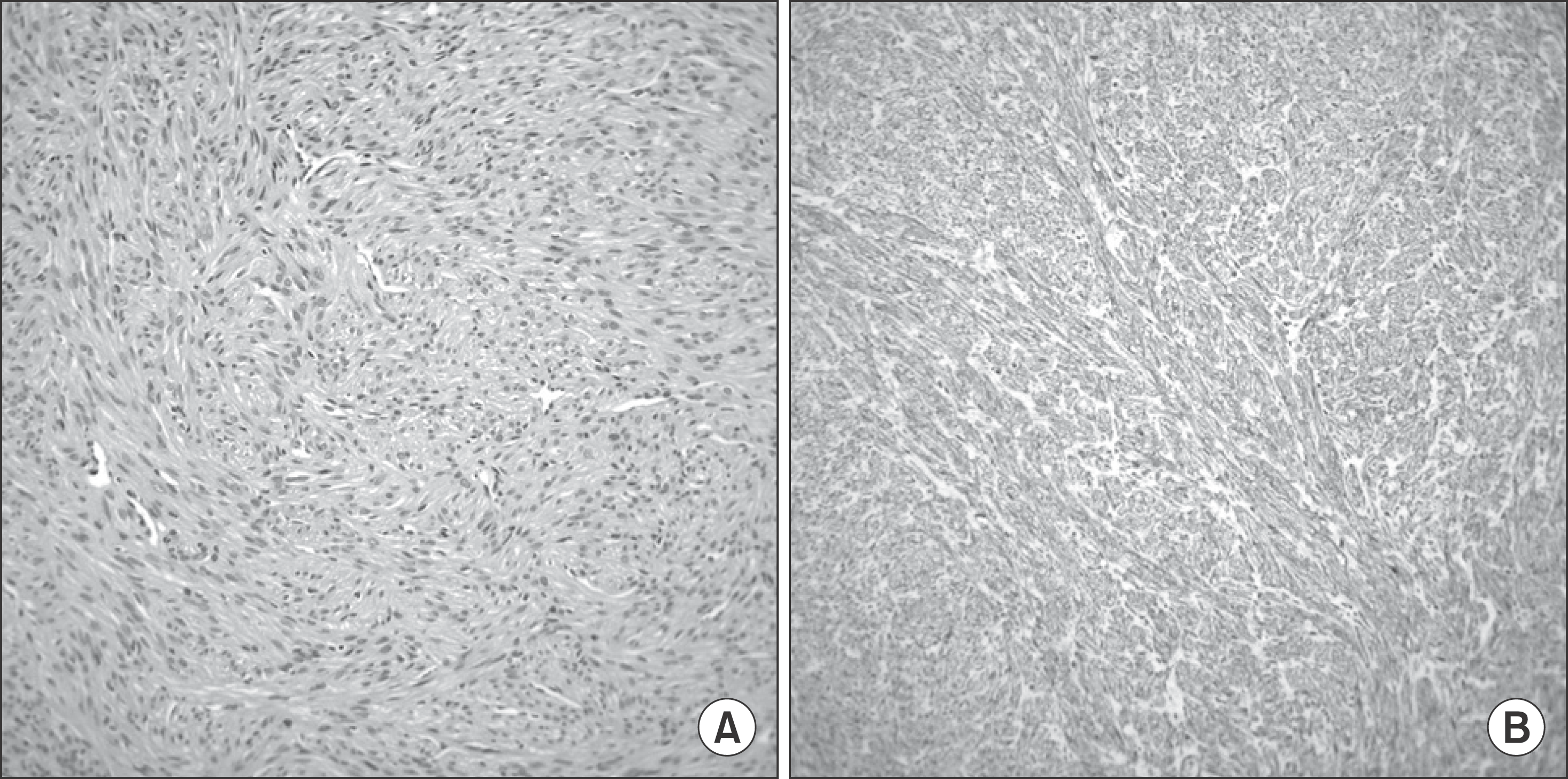
Figure 5.
Pathologic findings: (A) Tumor cells are composed of intersecting fascicles of well-differentiated smooth muscle cells that are characterized by a spindle shape, eosinophilic cytoplasm and blunt-ended, cigar-shaped nuclei (×200). (B) Smooth muscle actin is expressed in tumor cells (SMA, ×200).
 XML Download