

 PDF
PDF ePub
ePub Citation
 Print
Print


Abstract
Purpose
This study was aimed to evaluate the result of inlay cortical strut bone grafts for large cysts or cavitary bone lesions in long bones
Materials and Methods
Seven patients with large cyst or cavitary bony lesions were managed with curettage, allogeneic inlay cortical strut and cancellous bone grafts. Additional plate and screw fixations were performed in 6 patients. There were three SBCs, two FDs with secondary ABC changes, one FD and one post-cement spacer removal state. Three of them had pathologic fractures. Progression of bone healing and mechanical support and functional result were evaluated. The mean follow-up period was 25.4 months.
Results
Incorporations into host bones were progressed in all, average 4.2 months in six metaphyseal regions and 5.8 months in five diaphyseal regions respectively. Full structural supports were achieved in all except one patient without any additional procedures. No allograft-related complication was developed. Mean functional score according to the MSTS criteria was 29.6 at last follow up.
References
1. Killian JT, Wilkinson L, White S, Brassard M. Treatment of unicameral bone cyst with demineralized bone matrix. J Pediatr Orthop. 1998; 18:621–4.

2. Inoue O, Ibaraki K, Shimabukuro H, Shingaki Y. Packing with high-porosity hydroxyapatite cubes alone for the treatment of simple bone cyst. Clin Orthop. 1993; 293:287–92.
3. Leung PC, Chow YYN. Reconstruction of proximal femoral defects with a vascular-pedicled graft. J Bone Joint Surg Br. 1984; 66:32–7.
4. Sowa DT, Weiland AJ. Clinical applications of vascularized bone autografts. Orthop Clin North Am. 1987; 18:257–73.
5. Enneking WF, Dunham W, Gebhardt MC, Malawer M, Prichard DJ. A system for the functional evaluation of reconstructive procedures after surgical treatment of tumors of the musculoskeletal system. Clin Orthop. 1993; 286:241–6.
6. Mckay DW, Nason SS. Treatment of unicameral bone cysts by subtotal resection without grafts. J Bone Joint Surg Am. 1977; 59:515–9.
7. Oppenheim WL, Galleno H. Operative treatment versus steroid injection in the management of unicameral bone cysts. J Pediatr Orthop. 1984; 4:1–7.
8. Santori F, Ghera S, Castelli V. Treatment of solitary bone cysts with intramedullary nailing. Orthopedics. 1988; 11:873–8.
9. Fahey JJ, O'Brien ET. Subtotal resection and grafting in selected cases of solitary unicameral bone cyst. J Bone Joint Surg Am. 1973; 55:59–68.
10. Persson BM, Wouters HW. Curettage and acrylic cementation in surgery of giant cell tumors of bone. Clin Orthop. 1979; 120:125–33.
11. Byun YS, Shin DJ, Chang SA, Kwon DY. Inlay fibular autograft and helical LCP fixation for a segmental comminuted fracture of the osteoporotic proximal humerus. J Korean fracture Soc. 2006; 19:100–3.
12. Jung ST, Jeong KC, Kim HJ, Lee JH. Treatment of metaphyseal pathologic fractures of long bone using locking compression plate in chindren. J Korean Orthop Assoc. 2008; 43:539–43.
13. Cho DY, Koh ES, Lee MH. Operative treatment of proximal humeral fracture using inlay graft of fibular allograft combined with plating. J Korean fracture Soc. 1995; 8:84–92.
14. Mankin HJ, Gebhardt MC, Tomford WW. The use of frozen cadaveric allografts in the management of patients with bone tumors of the extremities. Orthop Clin North Am. 1987; 18:275–89.
15. Sim FH, Frassica FJ. Use of allografts following resection of tumors of the musculoskeletal system. Instr Course Lect. 1993; 42:405–13.
16. Glancy GL, Brugioni DJ, Eilert RE, Chang FM. Autograft versus allograft for benign lesions in children. Clin Orthop. 1991; 262:28–33.
17. Jaffe KA, Dunham WK. Treatment of benign lesions of the femoral head and neck. Clin Orthop. 1990; 257:134–7.
18. Stevenson S, Horowitz M. Current concepts review: The response to bone allografts. J Bone Joint Surg Am. 1992; 74:939–50.
Figure 1.
(A) An allogeneic ulna strut cortical bone and cancellous chip bone grafts. (B) After thorough curettage of bony lesion, inlay cortical strut and cancellous chip bones were grafted into the cavitary lesion and the plate and screw fixation was performed, in which the screws purchased the inlay cortical graft provided additional mechanical stability.
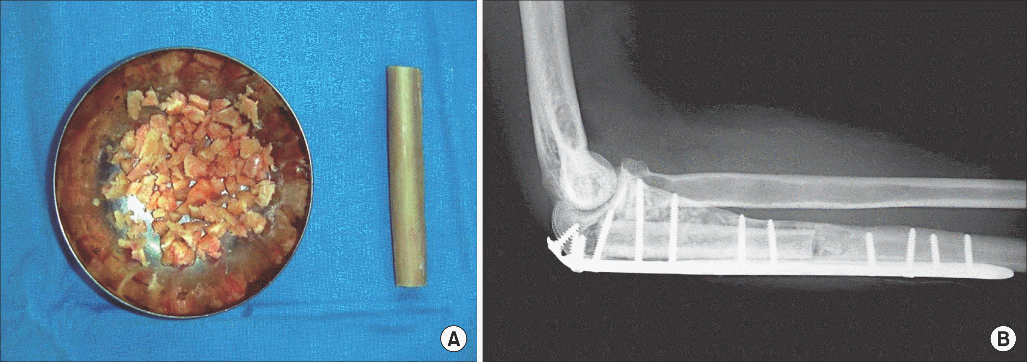
Figure 2.
(A) The lateral radiograph of 39 year old man revealed large cystic bony lesion in the proximal ulna with accompanied pathologic avulsion fracture of the olecranon. (B) In six month after operation, bone incorporation had occurred even at diaphyseal junction. (C) Post hardware-removal-radiograph demonstrated complete healing.
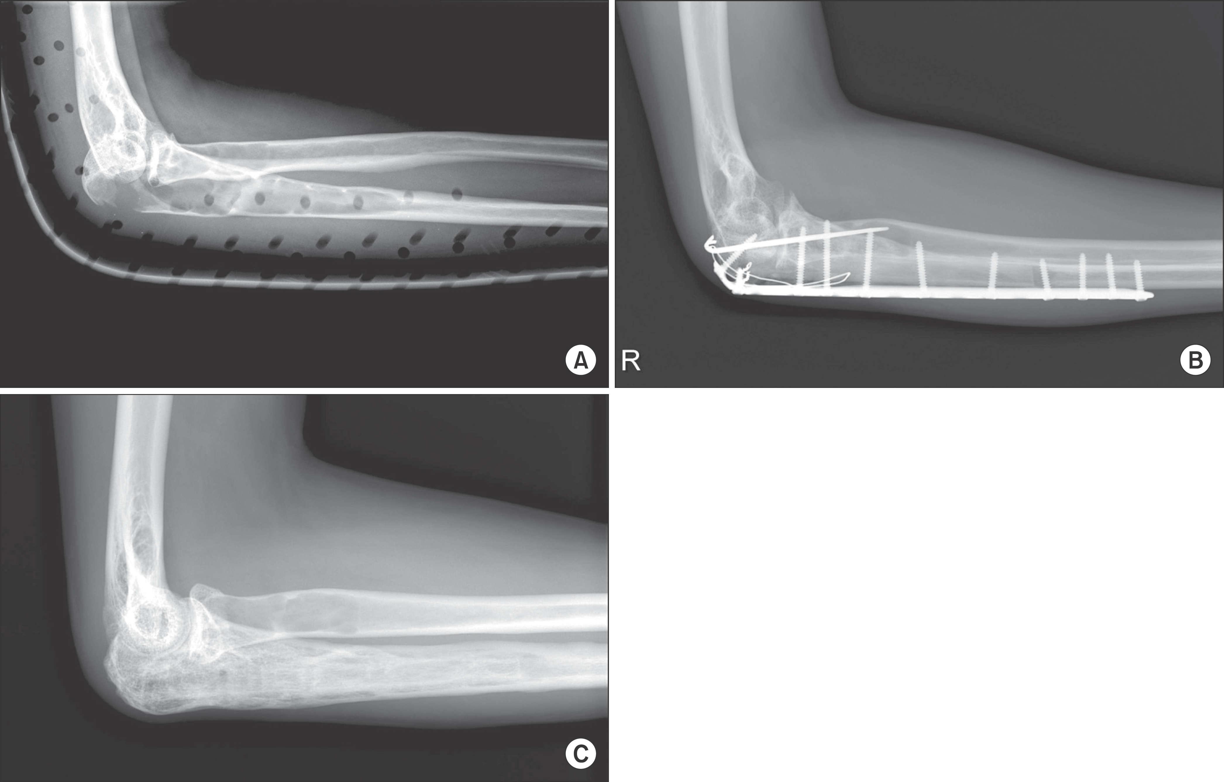
Figure 3.
(A) The 5 month postoperative radiograph of right humerus of 58 year old man revealed breakage of plate near the host-allograft bone junction with host bone fracture. (B) Complate incorporation and healing was obtained after a revisional plate fixation and autogenous iliac cancellous bone graft.
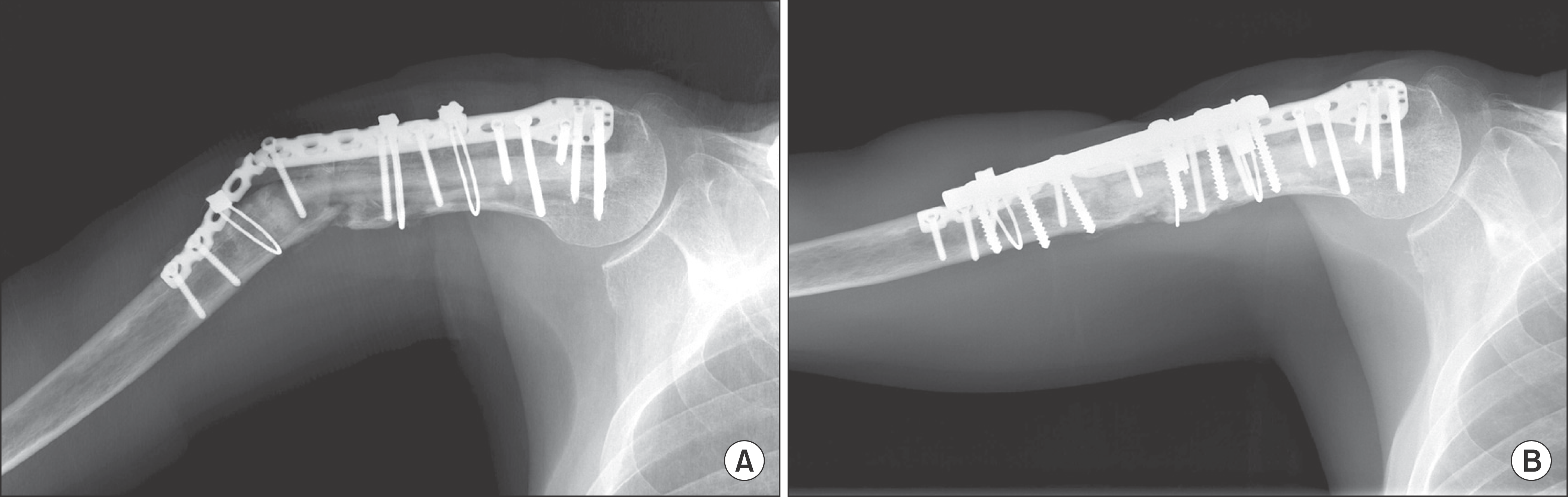
Figure 4.
(A) Both hip anteroposterior radiograph of 12 year old girl showed cystic bony lesion with pathologic fracture at right femur neck. (B) A 28 month follow-up scanogram revealed 14 mm leg length discrepancy due to early closure of growth plate at right proximal femur.

Table 1.
Summary of the Cases
 XML Download