Abstract
Purpose
We propose the measurement method of tumor movement for respiratory gated therapy in lung cancer patient for stereotactic radiosurgery, contouring method of tumor for radiation treatment planning using measured tumor movement. And through phantom study, we ascertain that the tumor movement is properly reflected in determination of PTV, and the tumor is properly and safely treated in full respiration phases and respiratory gated therapy.
Materials and Methods
Lung cancer phantom and 1-dimensional moving phantom were made to evaluate respiratory gated radiation therapy for lung SRS. 4D CT scan was performed using these phantoms and 10 sets of CT images and post-processed MIP (Maximum Intensity Projection) images were used to measure the tumor movement. The measured tumor movement in 4D CT images and MIP images were compared. Also, during radiation exposure in full respiration phases and respiratory gated phases, tumor movement included in radiation exposure was measured using EPID image and compared with measured data in 4D CT images and MIP images.
Results
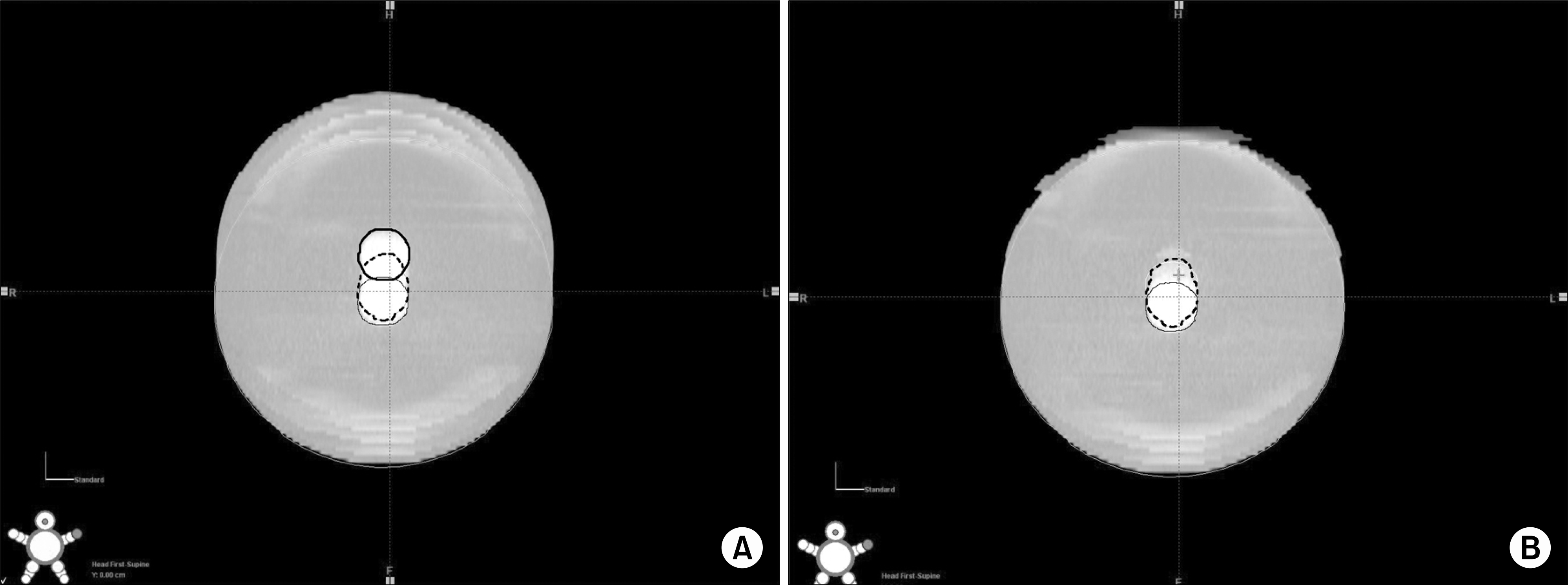
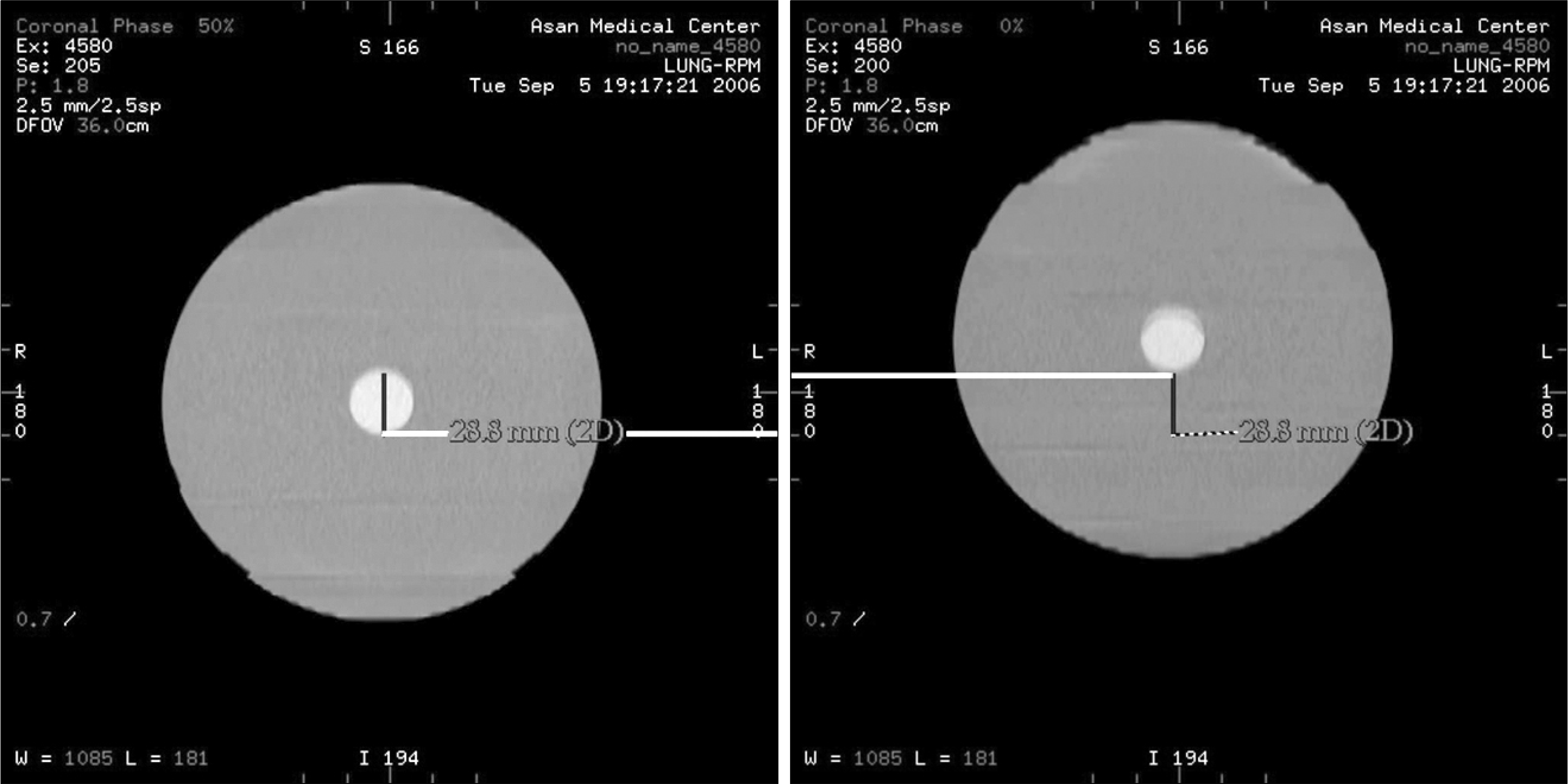
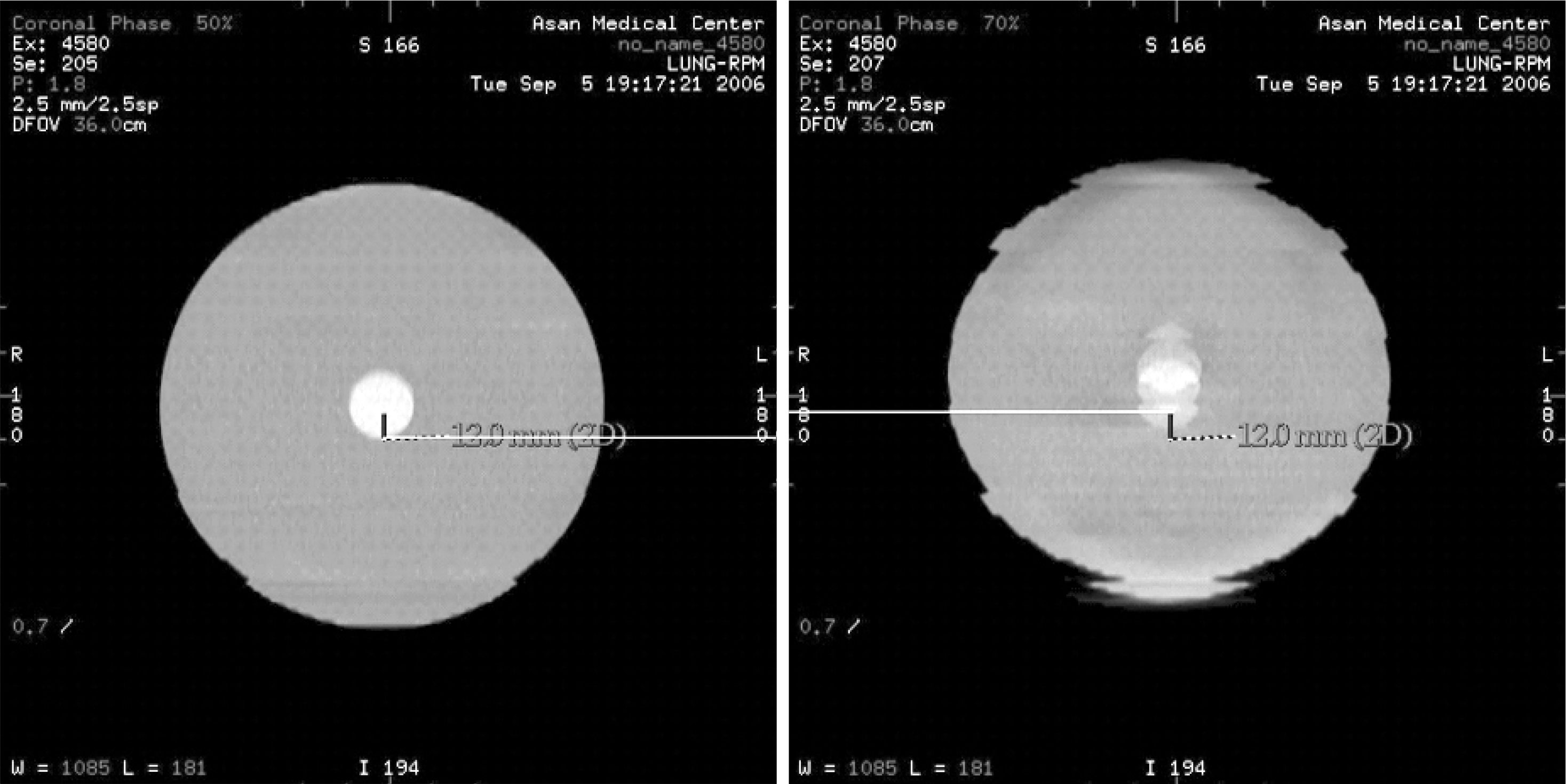
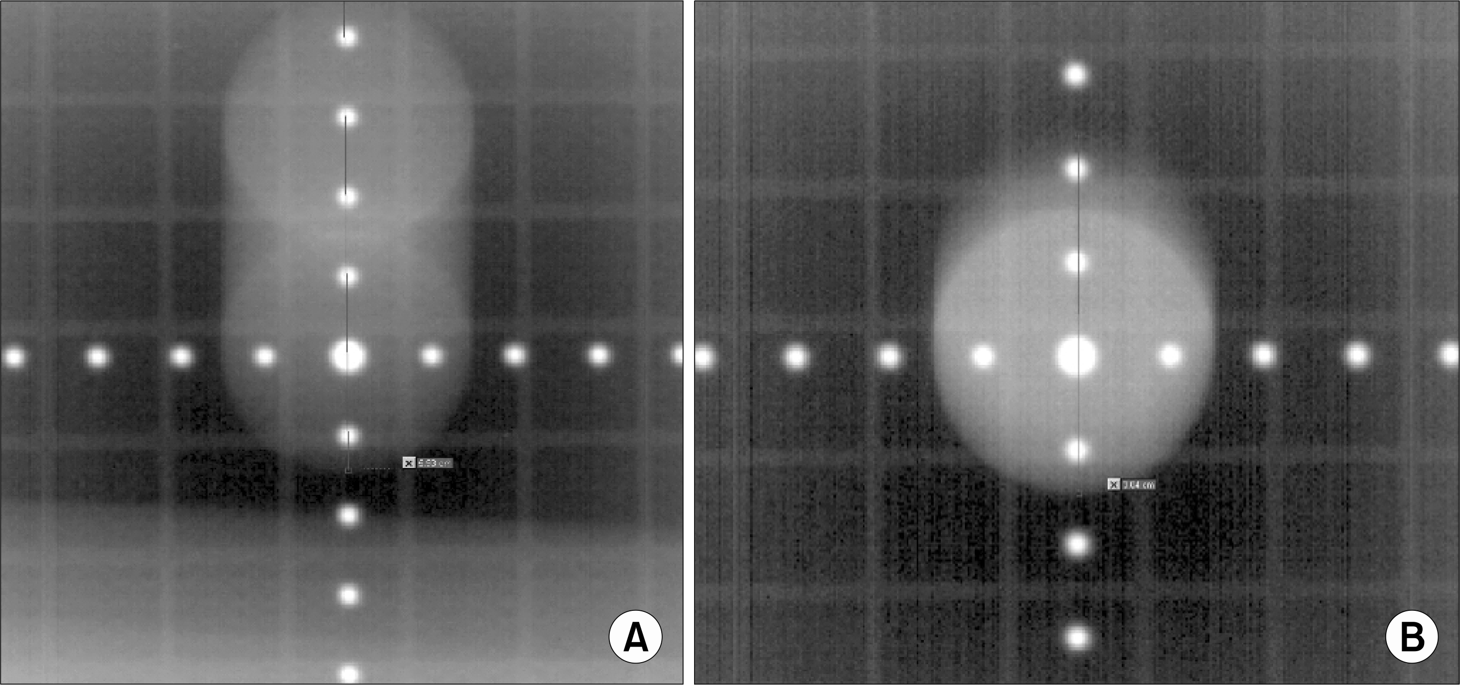
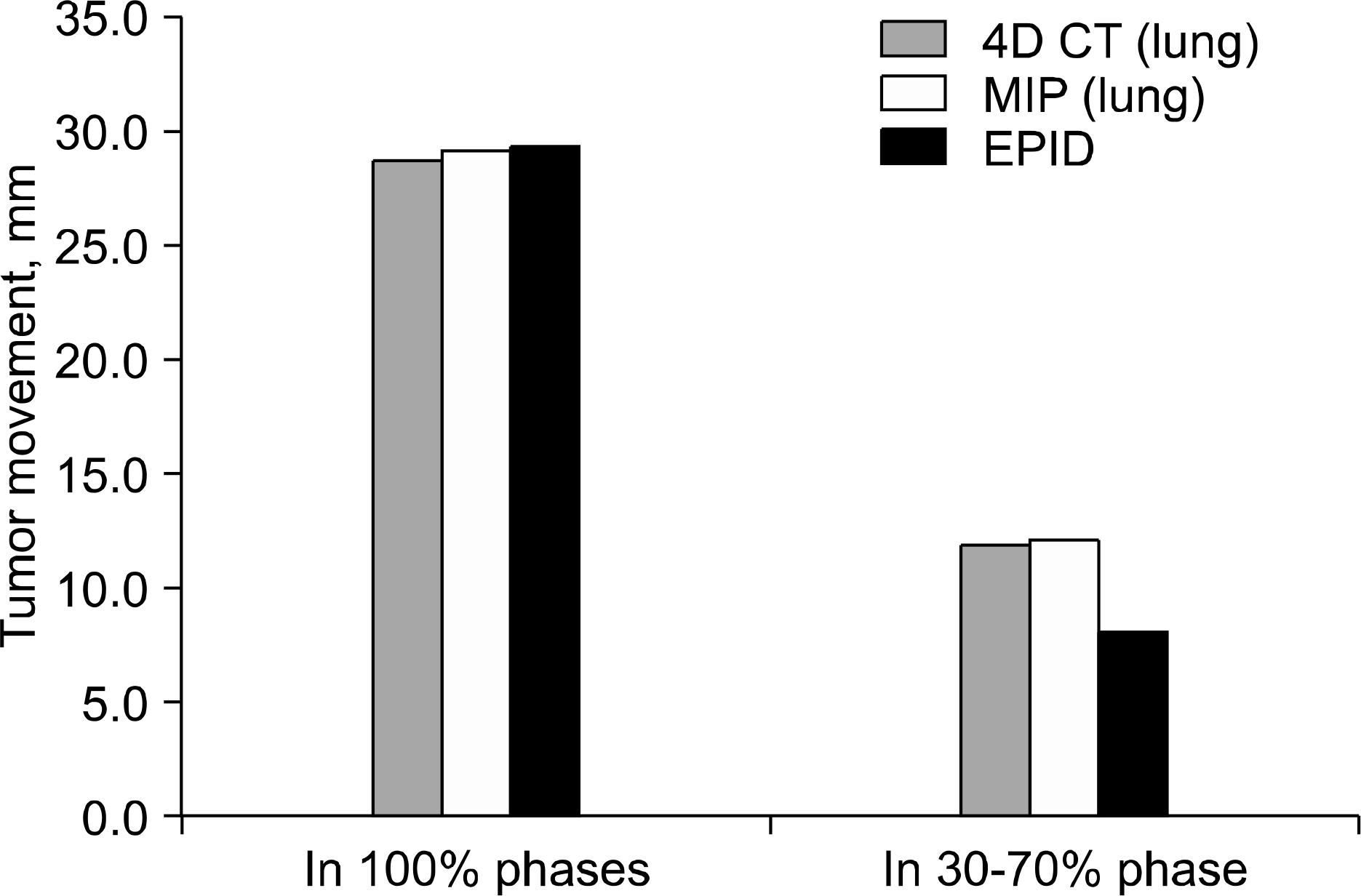
The tumor movement measured in full respiration phases was 28.8mm and 29.1 mm in 4D CT images and MIP images respectively, and in respiratory gated phases, 30∼70% phases, was 12 mm and 12.2 mm respectively. The tumor contoured in each phase images and MIP images was well agreed in full respiration phases and respiratory gated phases. The tumor movement included in radiation exposure was 29.3 mm and 8.4 mm in full respiration phases and respiratory gated phases respectively.
Conclusion
The tumor movement measured in 4D CT images and MIP images was well agreed, so we propose to use of MIP image for contouring of tumor in full respiration phases and respiratory gated phases. In full respiration radiation treatment and respiratory gated radiation therapy, the tumor movement included in radiation exposure was well agreed with measured tumor movement in 4D CT images or MIP images, so we ascertain though this phantom study we can exactly treat the tumor including tumor movement. In respiratory gated radiation therapy, the tumor movement included in radiation exposure was about 30% smaller than measured tumor movement in 4D CT images or MIP images, so we ascertain that we can safely treat the tumor including tumor movement in current provided technique.
References
1. ICRU Report 62. Prescribing, Recording, and Reporting Photon Beam Therapy (Supplement to ICRU Report 50), International Commission on Radiation Units and Measurements, Bethesda, MD (1999).
2. ICRU Report 50. Prescribing, Recording, and Reporting Photon Beam Therapy, International Commission on Radiation Units and Measurements, Bethesda, MD (1993).
3. McNair HA, Panakis N, Evans P, et al. Active Breathing Control (ABC) in Radical Radiotherapy of Non-small Cell Lung Cancer (NSCLC). Clin Oncol (R Coll Radiol). 2007; 19:S39.

4. Ko YE, Ahn SD, Yi BY, et al. Effectiveness of breath hold with a ABC for SRS of lung cancer. J Lung Cancer. 2005; 4:42–47.
5. D'Souza WD, Nazareth DP, Zhang B, et al. The use of gated and 4D CT imaging in planning for stereotactic body radiation therapy. Med Dosim. 2007; 32:92–101.
6. Wurm RE, Gum F, Erbel S, et al. Image guided respiratory gated hypofractionated stereotactic body radiation therapy (H-SBRT) for liver and lung tumors: initial experience. Acta Oncol. 2006; 45:881–889.

7. Schreibmann E, Chen GT, Xing L. Image interpolation in 4D CT using a BSpline deformable registration model. Int J Radiat Oncol Biol Phys. 2006; 64:1537–1550.

8. Shekhar R, Lei P, Castro-Pareja CR, et al. Automatic segmentation of phase-correlated CT scans through nonrigid image registration using geometrically regularized free-form deformation. Med Phys. 2007; 34:3054–3066.

9. van Herk M, Remeijer P, Rasch C, et al. The probability of correct target dosage: dose-population histograms for deriving treatment margins in radiotherapy. Int J Radiat Oncol Biol Phys. 2000; 47:1121–1135.

10. Jin L, Wang L, Li J, et al. Investigation of optimal beam margins for stereotactic radiotherapy of lung-cancer using Monte Carlo dose calculations. Phys Med Biol. 2007; 52:3549–3561.

11. Gordon JJ, Siebers JV. Convolution method and CTV-to-PTV margins for finite fractions and small systematic errors. Phys Med Biol. 2007; 52:1967–1990.

12. Jin JY, Ajlouni M, Chen Q, et al. A technique of using gated-CT images to determine internal target volume (ITV) for fractionated stereotactic lung radiotherapy. Radiother Oncol. 2006; 78:177–184.

13. Li XA, Qi XS, Pitterle M, et al. Interfractional variations in patient setup and anatomic change assessed by daily computed tomography. Int J Radiat Oncol Biol Phys. 2007; 68:581–591.

Fig. 2.
CT image of manufactured lung cancer phantom. It was seen that CT density of the tumor is larger than surrounding tissue.
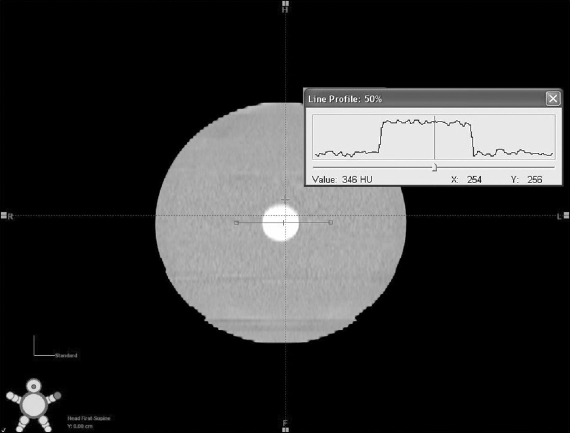
Fig. 5.
Tumor movement during (A) full respiratory phases and (B) respiratory gated phases (30∼70%) measured in MIP images.
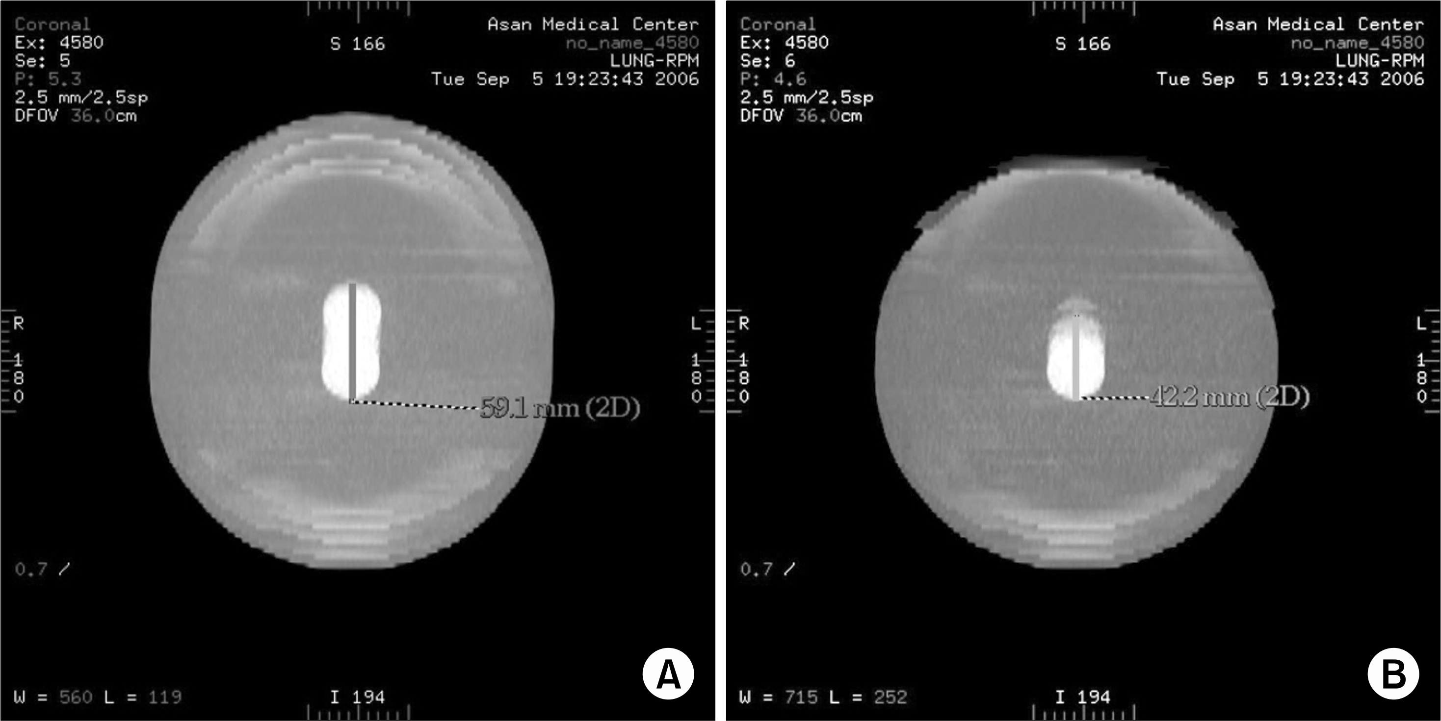
Fig. 6.
Tumor contoured in (A) CT images for every phase (thick solid for 0%, dotted line for 30%, solid line for 50%), (B) CT images for respiratory gated phases (dotted line for 30%, solid line for 50%) is well encompassed tumor shown in MIP image.


 PDF
PDF ePub
ePub Citation
 Print
Print


 XML Download