

 PDF
PDF ePub
ePub Citation
 Print
Print


Femoral shaft fracture is a commonly encountered injury. Intramedullary (IM) nailing is the preferred method for treating the fracture and viewed as the standard care for femoral shaft fractures.
There are three entry points for IM nailing: 1) the piriformis fossa,1) 2) the intercondylar notch,2) and 3) the greater trochanter.3) The piriformis fossa has the advantage of being colinear with the long axis of the femur, which reduces the risk of iatrogenic fracture comminution and varus malalignment.4) However, the disadvantages of this entry point are its sensitivity to anteroposterior translation5) and the technical difficulty. The intercondylar notch helps provide better fracture alignment of distal femoral shaft fractures,6) and the union rates achieved using modern techniques are similar to those of antegrade nailing. The disadvantages of this entry point include more complications related to the knee2) and relatively low union rates. The trochanteric entry point is technically easier due to subcutaneous location of the greater trochanter, especially in obese patients.7) Furthermore, it is less sensitive to anteroposterior translation due to more cancellous nature of trochanteric area, and it reduces the risk of iatrogenic bursting of the proximal segment.5) Its disadvantages are iatrogenic fracture comminution and varus malalignment.4) However, implants, which are specifically designed for trochanteric insertion with a proximal lateral bend and combined with the modified insertion technique, have been shown to essentially eliminate iatrogenic fracture comminution and varus malalignment.
We report on three cases of subtrochanteric femoral fracture during trochanteric femoral nailing for femoral shaft fractures. We here analyze the cases and surgical procedures, and include a review of the literature.
CASE REPORTS
The authors oversaw three cases of iatrogenic subtrochanteric femoral fracture during trochanteric femoral nailing for femoral shaft fractures, between April 1, 2009 and May 31, 2009. Each case was treated by a different surgeon. There were no abnormal bowing of the contra-lateral femurs in the preoperative radiographs. The trochanteric IM nail used in all cases was the Sirus nail (Intramedullary Nail System, Zimmer, Cowpens, SC, USA). Based on the manufacturer's recommendations, all surgical procedures were conducted as follows. Using an entry point directly lateral to the tip of the greater trochanter, the intramedullary canal 1 mm larger than the chosen nail diameter was reamed. The IM nail was inserted over the guide wire while anteriorly orientating the targeting device, and the targeting device was slowly rotated by pushing the nail further down the intramedullary canal after passing the proximal metaphysis. After fully inserting the nail, the targeting device was rotated by approximately 90°.
Case 1
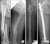
A 27-year-old woman (height 157 cm and weight 48 kg), with chronic renal failure and a previous complete lumbar spinal cord injury, sustained a femoral shaft fracture (Arbeitsgemeinschaft für Osteosynthesefragen/Orthopaedic Trauma Association [AO/OTA] classification 32B1) (Fig. 1A). After reaming up to 13 mm, a 12 mm diameter, and 340 mm long nail was introduced manually. During the passage of the proximal metaphysis, resistance was felt. We then checked for evidence of fracture using an image intensifier, but no fracture was detected. Insertion of the IM nail was progressed with the image intensifier, focused around the fracture site. After inserting the nail, and while attempting to fix the proximal locking screw, a 7-8 cm long longitudinal fracture was observed along the lateral cortex from the vastus ridge to below the lesser trochanter. Due to severe osteoporosis (T score, -4.3), we believed that poor bone quality had caused the iatrogenic subtrochanteric fracture in this patient (Fig. 1B and C).
Case 2
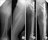
A 63-year-old man (height 169 cm and weight 72 kg) with multiple trauma injuries presented with a middle femoral shaft fracture (AO/OTA classification 32B1) (Fig. 2A). After reaming up to 14 mm, a 13 mm diameter, and 360 mm long nail was inserted. However, when the targeting device was slowly rotated following the passage of the proximal metaphysis after confirming the central direction of nail's tip during insertion, resistance was felt, and cortical breakage along the lateral cortex in the subtrochanteric area was observed under an image intensifier. The newly developed fracture was on the lateral side and had a distance from the original fracture site; therefore, we considered it as a missed, concealed ipsilateral proximal femoral fracture rather than an iatrogenic fracture comminution. Accordingly, we continued the nail insertion (with gradual rotation) until a cracking sound was heard. The result was a displaced complete subtrochanteric fracture (Fig. 2B and C).
Case 3
A 70-year-old man (height 160 cm and weight 63 kg) sustained a proximal femoral shaft fracture (AO/OTA classification 32A1) in a traffic accident (Fig. 3A). No comminution was observed around the fracture surfaces. After reaming up to 12 mm, an 11 mm diameter, and 320 mm long nail was introduced manually. After the nail passed the proximal metaphysis, it was derotated gradually with successive mallet blows until it reached its intended location. When the proximal metaphysis was checked prior to proximal locking screw fixation, a newly developed proximal femoral fracture was observed under an image intensifier. This fracture started from the previous proximal fracture surface in the lateral aspect and propagated almost to the lesser trochanter, resulting in a proximal femoral shaft fracture with subtrochanteric extension (Fig. 3B and C).
DISCUSSION
Trochanteric IM nails with anatomical ante curvature can easily be inserted in cases of femoral shaft fractures with excessive anterior bowing, which are frequently encountered in Asia.8) One of the concerns of the trochanteric IM nailing is comminution or angulation of the intact proximal femoral fragment.3) Varus malalignment and iatrogenic fracture comminution were previously associated with the use of straight nails inserted through this entry portal.9) They were related to a medially directed insertion angle which were occurred during the insertion of straight nails.4) Implants, specifically designed for trochanteric insertion with a proximal lateral bend, in combination with the modified nailing technique, have been shown to essentially eliminate varus malalignment and iatrogenic comminution.3,9) Rotation of the nail by 90° upon insertion, such that the anterior bow is apex medial and the direction of the tip of the nail is central with gradual derotation after the nail crosses the fracture, is the main modification adopted in the modified nailing technique as compared with the standard nailing technique.4)
We analyzed our cases to identify the cause of iatrogenic subtrochanteric femoral fractures and thoroughly reviewed the surgical procedures. First, we noted on the proper timing of derotation. The manufacturer's recommendation on the timing of derotation is as follows: "After passing the proximal metaphysis, the targeting device is slowly rotated by pushing the nail further down the intramedullary canal with successive mallet blows." However, before the nail passes the fracture site, derotation itself causes more hoop stress in the subtrochanteric area due to continued anterior bowing of the nail. Moreover, given the less cancellous nature of the subtrochanteric area, it is less forgiving of hoop stress; therefore, this action may increase the risk of iatrogenic bursting in the subtrochanteric area. As has been reported, the targeting device must be derotated gradually with successive mallet blows after the nail crosses the fracture.4) Second, the subtrochanteric region has several unique characteristics. The femoral canal in the subtrochanteric region has a tubular shape and a wider mediolateral dimension as compared with the anteroposterior dimension. The subtrochanteric area is mainly composed of cortical bone, giving its less cancellous nature, and is more unforgiving than the trochanteric area with regard to generated hoop stress. Furthermore, large biomechanical stresses are present in the subtrochanteric area under loaded condition. In addition, a dense vertical plate of bone, calcar femorale, extends from the posterior medial portion in the subtrochanteric area and is the thickest medially. Third, modern reaming techniques call for a minimum (0.5 to 1 mm) of reaming beyond the occurrence of cortical chatter at the level of the isthmus, and then the proper nail diameter for a snug fit is recommended as 1 to 1.5 mm smaller than the largest reamer used.6) Rotation of the nail by 90° upon insertion in the trochanteric nailing technique is an important modification of the standard nailing technique, originally introduced to avoid iatrogenic comminution. This rotation is performed, so that the anterior bow of the nail is apex medial, and the tip of the nail is directed centrally.6) Due to wider dimension of mediolateral plane in the subtrochanteric region, with the rotation of the nail by 90°, it is not difficult to pass the nail through the subtrochanteric region. On the other hand, derotation with successive mallet blows after the nail crosses the fracture may abut the nail to the anterior cortex of subtrochanter and generate excessive hoop stress at the lateral cortex. Such is likely to break first due to its comparative weak anatomic and biomechanical properties. In all cases, subtrochanteric fractures occurred in the lateral area alone (Case 1) or started from the lateral area (Cases 2 and 3), showing that these fractures propagated from the lateral side due to the anterior nail bow causing lateral pressure when rotated for trochanteric entry. Because of the ante curvature of the trochanteric nail and its anterior bowing in proportion to its length, the effective nail diameter increases during derotation, and subsequently develops a mismatch between nail diameter and subtrochanteric canal diameter. Therefore, the manufacturer's recommendation that a nail 1 mm smaller than the largest reamer used becomes inadequate. If serious resistance happens during nail passage, the impaction must be stopped, and the cause should be determined using an image intensifier. If necessary, a nail with a small diameter must be used, or the medullary canal must be reamed to a larger diameter. This undersizing of the nail is required to avoid iatrogenic subtrochanteric femoral fractures. Fourth, the manufacturer recommends that the trochanteric nail entry point be lateral to the tip of greater trochanter. It must be noted that the very tip of the trochanter is not necessarily the proper starting point for trochanteric IM nailing in every patient. There is anatomic variation with regard to the alignment of the tip of the trochanter, relative to the long axis of the femoral shaft.10) Thus, as reported previously, the proper entry point for trochanteric nailing is just lateral to the long axis of the femur.4) As such, the precise proper starting point for trochanteric nailing is individualized for each patient; but it is usually at slightly medial to or slightly lateral to the tip of the greater trochanter. Furthermore, if lateralization of the trochanteric mass is observed, the nail is not inserted at the apex of the greater trochanter, but rather at the base of the femoral neck to restore nail alignment with the femoral diaphysis. We suspect the main reason for the iatrogenic fracture in Case 3 was the wrong entry portal. In this case, the portal was made too laterally which led the nail direction into varus, resulting in medial comminution. This complication takes place frequently, because the entry point is made with inadequate control of the proximal fragment. We consider that this mismatched nail entry points generated undue force during nail insertion and contributed to the iatrogenic subtrochanteric femoral fractures. Lastly, it may be that the anatomical contour of the nail is not suited to the Korean population, as there have been no abnormal bowing of the contra-lateral femurs in the preoperative radiographs.
The trochanteric IM nail is a useful implant for treating femoral shaft fractures. The following surgical procedures should be considered for the trochanteric nailing of femoral shaft fractures to prevent implant-derived complications. First, the proper starting point for trochanteric nailing is just lateral to the long axis of the femur. Second, nail derotation must be performed after the nail crosses the fracture site completely. Third, when resistance is encountered during the nail passage, the procedure must be stopped, and the cause should be determined using an image intensifier. If necessary, a nail with a smaller diameter must be used or further reaming performed, rather than continuing after screening for a fracture.
 XML Download