

 PDF
PDF ePub
ePub Citation
 Print
Print


Ulnar nerve injury subsequent to a fracture of the distal radius is extremely rare compared to a median nerve injury, which has an incidence of 2-7%.1-3) Bacorn and Kurtzke4) reported only 1 case of ulnar nerve injury (0.05%) in 2,000 patients with a fracture of the distal radius. Ulnar nerve injuries following a fracture of the distal radius can be caused by traction or contusion by bone fragment, compression by fibsosis or swelling of the adjacent tissues, intraneural fibrosis, and laceration.3,5-7) This case report presents two cases of ulnar nerve palsy after a closed fracture of the distal radius. Their neurological symptoms sustained after fixation of the fracture recovered with nerve decompression and neurolysis at 2 or 3 months after the trauma.
CASE REPORTS
Case 1
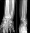
A 19-year-old male motorcycle rider was hit by a car and complained of pain in the left wrist. A physical examination revealed tenderness of the left wrist and hypesthesia of the 4th and 5th finger. The plain radiographs revealed an intraarticular comminuted fracture of the distal radius with posterolateral displacement of the distal fragment and an ulnar styloid process fracture (Fig. 1).
A closed reduction and percutaneous K-wires fixation with an external fixator were performed on the 2nd day after the trauma. Postoperatively, the patient complained of a continuous tingling sensation at the medial 1/2 of the 4th and 5th fingers and the progressively development of a clawing deformity. At 6 weeks after surgery, the external fixator was removed and the range of motion exercise was started. At 8 weeks postoperatively, ultrasonography and electrophysiologic study were performed due to the continuous ulnar nerve palsy. The ultrasonographic findings showed that the continuity of the ulnar nerve was maintained but there was swelling of the ulnar nerve in the Guyon's canal and fibrosis of the tissues around the nerve. The electrophysiologic study revealed a decrease in the sensory and motor conduction velocity of the ulnar nerve and denervation potential in the intrinsic muscle of the hand.
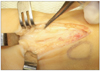
At 12 weeks postoperatively, ulnar nerve exploration, decompression and neurolysis was performed due to the lack of improvement in the neurological symptoms. The intraoperative findings revealed swelling of the ulnar nerve and compression by the adjacent fibrous tissues and adhesion in the Guyon's canal (Fig. 2). At the 3rd month since the nerve exploration, the ulnar nerve palsy had recovered completely.
Case 2
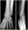
A 27-year-old male complained of pain in his left wrist after a fall from a height. A physical examination revealed severe swelling and tenderness in the left wrist and hypesthesia in the 4th and 5th finger were observed. The plain radiographs revealed a severe intraarticular comminuted fracture of the left distal radius with an ulnar styloid process fracture (Fig. 3).
An open reduction was performed on the day of the trauma. A small 2 cm-incision was made on the voloradial side to expose the fracture site. A reduction of the articular surface was attempted using a periosteal elevator. The bone deficiency in the metaphysis was treated with an allogeneous cancellous bone graft, and an external fixator was applied. Postoperatively, the patient complained of a tingling sensation in the medial 1/2 of the 4th and 5th finger and clawhand deformity. The ulnar nerve palsy was still present 6 weeks after surgery, which required ultrasonography and an electrophysiologic study. The ultrasonographic findings showed that the continuity of the ulnar nerve was maintained but there was swelling of the nerve in the Guyon's canal. The electrophysiologic study revealed a decrease in the sensory and motor conduction velocity of the ulnar nerve and the denervation potential in the intrinsic muscle of the hand.
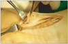
At 8 weeks postoperatively, ulner nerve exploration, decompression and neurolysis was performed due to the lack of improvement in the neurological symptoms. The intraoperative findings revealed swelling of the ulnar nerve and adhesion by the adjacent fibrous tissues (Fig. 4). The neurological symptoms began to improve at the 4th week since the nerve exploration. At 1 year postoperatively, minor numbness was felt in the 4th finger and 5th finger but the clawhand deformity had disappeared.
DISCUSSION
A median nerve injury following a fracture of the distal radius is a relatively common neurological complication that is found in 2-7% of cases and is associated with high energy trauma. Carpal tunnel syndrome after a fracture of the distal radius is caused by an increase in pressure on the carpal tunnel due to swelling or bleeding into the carpal tunnel in most cases.1-3) In contrast, ulnar nerve injury is an extremely rare event with only 30 cases reported worldwide.2-10) According to Bacorn and Kurtzke4) an ulnar nerve injury was found in only 1 out of 2,000 patients (0.05%) with a fracture of the distal radius. However, Soong and Ring3) suggested a higher frequency of ulnar nerve injuries, showing that 5 of their study population treated for a distal radial fracture within a 2 year period presented with an ulnar nerve injury. In our experience, the incidence of ulnar nerve injury was similar to that reported by Soong and Ring3). We encountered 4 cases of ulnar nerve injury subsequent to a fracture of the distal radius. Two of them were combined with a closed fracture and 2 of them were accompanied with open fracture involving severe soft tissue damage, such as a degloving injury. We postulate that the higher incidence of severe displacement, comminuted fractures, intra-articular fractures and open fractures are due to an increase in the number of high-energy injuries caused by motor vehicle accidents and industrial accidents.
The cases reported thus far have also shown that an ulnar nerve injury mostly affects young people as a result of a high-energy injury caused by traffic accidents, falls from a height and sports injuries, and is common in patients with severe displacement and comminution, combined a distal ulnar fracture and open fracture. The 2 young patients in this study resulted from a high-energy injury caused by a motorcycle accident and a fall from a height. In them, the displacement or intra-articular comminution was severe and combined with an ulnar styloid process fracture.
An ulnar nerve injury is caused primarily by direct contusion, traction and nerve compression due to fibrosis of the adjacent tissues or swelling, intraneural fibrosis, and rarely by laceration in a distal radial fracture.3,5-7) Zoega7) reported from their intraoperative findings and a cadaver study that an ulnar nerve injury can occur as a result of contusion caused by a posterior and radial displacement of the distal radius fragment. Clark and Spencer5) reported that compression by thick fibrous tissues around the ulnar nerve resulted in progressive ulnar nerve palsy and demonstrated that a permanent ulnar nerve injury could be avoided even when it was displaced or extended in a fracture of the distal radius because it has a higher mobility and extensibility than the median nerve. According to Soong and Ring3) the ulnar nerve is more vulnerable to traction and contusion than to compression because the ulnar nerve is located outside the carpal tunnel and is fixed in the Guyon's canal. Considering that our cases presented with swelling as a result of a contusion of the ulnar nerve as well as compression and adhesion by the adjacent tissues, it is believed that traction, contusion and compression by the adjacent fibrous tissues are the main causes of an ulnar nerve injury in a closed fracture with laceration being a rare cause. However, it is not believed that an ulnar nerve injury is caused by an open fracture involving extensive damage to the soft tissues.
With regard to the treatment of an ulnar nerve injury after a distal radial fracture, there is no disagreement regarding the need for early nerve exploration when combined with an open fracture or acute carpal tunnel syndrome. However, there is some controversy regarding the treatment of an ulnar nerve injury in a closed fracture. Some authors reported that careful observation can lead to recovery,3,7) while Vance and Gelberman6) reported that rapid recovery could be obtained with early decompression. In addition, Bourrel and Ferro1) suggested that neurolysis should be performed when there are no signs of neural recovery in the 6 months after the trauma. In our cases, the neurological symptoms did not disappear after reduction and fixation of the fracture, and no recovery was observed after 2-3 months of trauma. Ultrasonography and an electrophysiologic study were performed, and the location and pattern of the ulnar nerve injury were observed. Nerve decompression and neurolysis led to successful outcomes.
In conclusion, an ulnar nerve injury following a fracture of the distal radius is rare. However, median and ulnar nerve injuries should be considered when a high-energy injury, wide displacement, or comminution is accompanied. Considering that an ulnar nerve injury combined with a closed fracture of the distal radius results from a contusion or compression by the adjacent tissues in most cases, the treatment options should be chosen depending on the degree of recovery after an anatomical reduction and fixation of the fracture. When signs of recovery are not observed during the observations, nerve exploration after ultrasonography and an electrophysiologic study is recommended to identify the pattern, location and cause of the injury as well as the possibility of regeneration.
 XML Download