

 PDF
PDF ePub
ePub Citation
 Print
Print


Dear Editor:
Anetoderma is a rare cutaneous disorder characterized by localized depressions or the outpouchings of the skin caused by laxity and the weakening of the dermal connective tissue as a result of the focal loss of elastic fibers1. Anetoderma has two forms: primary and secondary. Primary anetoderma develops on clinically normal skin without any preceding dermatoses. Secondary anetoderma develops on exactly the same area as a previous specific pathology1.
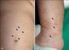
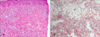
A 3-month-old girl presented with an increasing number of multiple atrophic and brownish scaly macules on her trunk and both extremities, which had begun to develop 3 weeks previously. She had erythematous asymptomatic papules that changed to brownish scaly macules. On clinical examination, the otherwise healthy-looking girl was presented with 40 to 50 atrophic and round-to-oval shaped brownish macules on her trunk and both extremities (Fig. 1). The lesions were 2 to 10 mm in diameter and were relatively well circumscribed, presenting as depressions below the level of the normal skin. A biopsy was taken from a typical-looking lesion on her leg. Since the clinical features including the lesions and the patient's age closely resembled primary anetoderma and looked just like a case we had previously reported, we presumed that this was a case of primary anetoderma. However, a histopathological examination revealed degenerated collagen bundles along with histiocytes, lymphocytes and Touton giant cells in the dermis (Fig. 2A). An alcian blue staining was positive in the degenerated collagen fibers.
The diagnosis was therefore granuloma annulare. The elastic stain also revealed a complete elastolysis or fragmented elastic fibers in the superficial and the mid dermis (Fig. 2B). These findings were consistent with anetoderma. Thus, histopathologic features of granuloma annulare and anetoderma coexisted in this patient.
Anetoderma occurs mainly in women aged 20 to 40 years, but is occasionally reported in younger and older patients of both sexes1. It has also occasionally been described/reported in premature infants, which may be related to the use of cutaneous monitoring leads or adhesives1.
Granuloma annulare generally resolves spontaneously without leaving any atrophic scars2. However, there are some reported cases manifesting the development of mid-dermal elastolysis and anetoderma following granuloma annulare3,4. Güneş et al.2 found that elastic fiber damage is one of the main accompanying features of granuloma annulare, which may develop from delayed type hypersensitivity. Thus, elastic fiber destruction may be intrinsic to granuloma annulare and can be severe enough to cause visible skin atrophy like anetoderma2,4. In addition, elastolytic enzymes released from inflammatory cells are thought to be responsible for the fragmentation of elastic fibers. Vibronectin and decay acceleration factor have also been implicated5.
Anetoderma should be distinguished from several elastolytic disorders including acquired cutis laxa, mid-dermal elastolysis, and perifollicular elastolysis4. Acquired cutis laxa differs from anetoderma by its generally widespread sagging or lax skin. Mid-dermal elastolysis is characterized by wrinkly patches and follicular papules, and the selective loss of elastic fibers limited to the mid dermis. Perifollicular elastolysis are presented as small papules centered around the follicles on the face or the upper back.
We report an interesting case of anetoderma developing in generalized granuloma annulare, with the earliest known age of onset and displaying coexisting histopathologic features of granuloma annulare and anetoderma.
 XML Download