

 PDF
PDF ePub
ePub Citation
 Print
Print


Dear Editor:
Cryptococcosis is an unusual opportunistic infection, and is characterized by a high mortality rate, especially among those with cryptococcaemia1. Cryptococcal skin lesions are often polymorphous in appearance. However, widespread cutaneous lesions and necrotizing fasciitis (NF) have rarely been reported. Here, we present a case of disseminated cryptococcosis with widespread cutaneous lesions, NF and cryptococcemia, occurring in an immunosuppressed patient.
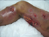
A 41-year-old woman was transferred to our hospital, with a history of skin necrosis on the right lower limb and high fever for 17 days, and erythemas, indurations, and abscesses on the other three limbs for 14 days. She was diagnosed as NF in another hospital, and treated with meropenem empirically, but without noticeable effect. She suffered from chronic glomerulonephritis, chronic renal insufficiency, and was receiving maintenance immunosuppressive therapy with prednisone (30 mg/d).
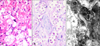
On examination, black necrotic skin, bare subcutaneous tissue and multiple abscesses, mainly situated on the right leg, were the most obvious clinical manifestation (Fig. 1). Multiple erythemas, indurations and abscesses could be found on the other three limbs. Chest x-ray demonstrated bilateral infiltration in the lung, with pleural effusion on the right. Incision and drainage of the abscess were performed immediately, and thorough surgical debridement was performed 3 days later. On the 7th hospital day, pathological examination reported encapsulated yeast in subcutaneous tissue, suggesting Cryptococcus neoformans (Fig. 2). This was also isolated and identified (VITEK 2-compact) from blood, and the wound. A follow-up culture of cerebrospinal fluid was negative. When the definite diagnosis of disseminated cryptococcosis, cryptococcemia, cryptococcal NF, bilateral pulmonary infection and renal failure had been established, intravenous fluconazole (400 mg daily) was initiated. This dosage of fluconazole was continued for one month, and was then reduced by half, for another month. An oral maintenance dose was then given, for a further 4 months. She was discharged 54 days after admission, with her wounds completely closed with razor-thickness skin grafts.
Cutaneous dissemination occurs in approximately 10% of cases with cryptococcosis2. Most patients with cutaneous involvement have lesions, consisting of ecchymosis, papules, nodules, vesicles, ulcers, abscesses, and very rarely, NF3,4. The patients with cryptococcal NF are usually treated empirically for a presumed bacterial infection, without response, as in the present case.
C. neoformans can be isolated from blood cultures in 10% to 30% of patients with cryptococcosis1. Previous studies of patients with cryptococcemia have shown a high mortality rate, which was reported to be between 31% and 41%1,5. Cryptococcaemia signifies a fulminant form of cryptococcal disease, and requires early diagnosis, and prompt antifungal therapy.
Antifungal therapy is the cornerstone to treat disseminated cryptococcosis and cryptococcemia. However, for NF, and serious, large areas of cutaneous infections, surgical treatment is indispensable. Thorough debridement and drainage, reducing the chance of hematogenous spread, and preventing muscle necrosis, sepsis and death, were one of the keys to cure the patient.
This report highlights the need to recognize this rare infectious disease and its clinical manifestations, especially among those patients who are receiving treatments that produce a state of immunosuppression.
 XML Download