

 PDF
PDF ePub
ePub Citation
 Print
Print


Dear Editor:
The frequent presence of Candida albicans in chronic paronychia is well known. Patients with chronic mucocutaneous candidiasis, involving the nail plate and proximal nail fold, can show chronic paronychia with a pseudo-clubbing or 'chicken drumstick' appearance1. Here, we described a case of chronic paronychia without any Candida species involved, but complicated by onychomycosis, caused by Trichophyton rubrum.
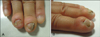
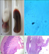
A 46-year-old woman presented with a 3-year history of swelling of the right hand's third and fourth nail folds, and a 1-year history of nails alteration. The patient stated that the dermatoses had never been treated, and there was no history of trauma, or family onchoymycosis. Dermatological examination showed that most nails of the involved fingers were lost (Fig. 1A), and the proximal and lateral nail folds were obviously swollen, and developed a pseudo-clubbing (Fig. 1B). No other nails and skin were affected. The woman was otherwise healthy. Direct microscopic examination of nail clippings revealed branching fungal hyphae. Culture growth and slide culture showed T. rubrum (Fig. 2A, B). Those of the nail folds were negative. Histopathological examination showed squamous cell hyperplasia, with parakeratosis and chronic inflammation in the dermis (Fig. 2C, D). The radiograph of fingers revealed soft tissue swelling, and no abnormality of the bones. A diagnosis of chronic paronychia, complicated by onychomycosis caused by T. rubrum, was made. Itraconazole 200 mg twice daily for one week, and topical triamcinolone acetonide acetate cream 0.05% on the nail folds twice daily for one month, were prescribed. Mycological examination after one month was negative, but there was no obvious progress of the swelling lesions, and no new nail grown out. She was disappointed, and subsequently lost to follow-up.
Chronic paronychia is an inflammatory disorder of the proximal nail fold, reactive to irritants and allergens2,3,4. When the cuticle separates from the nail plate, the region between the proximal nail fold and nail plate is vulnerable to infection by bacterial and fungal pathogens3. Colonization with C. albicans or bacteria may occur in the lesion2,4. Paronychia is associated with 84.2% of candidal onychomycosis; the initial paronychia leads to nail discoloration, and then, dystrophy ensues4. Patients with chronic mucocutaneous candidiasis involving the nail plate and proximal nail fold can show swelling of the proximal nail fold, with considerable expansion of the terminal phalanx, and develop a pseudo-clubbing, or 'chicken drumstick' appearance1,5.
In our case, dermatophyte onychomycosis may be the result of chronic paronychia, which destroyed the skin barrier, altered the quality of nail, and led to nail loss.
Treatment with antifungal agents was effective, and the patient quickly achieved mycological cure, after oral itraconazole pulse therapy for 1 month. This may because a little nail was preserved, and the fungal load was low.
Our case presents an appearance of pseudo-clubbing, which is one feature of chronic mucocutaneous candidiasis, but with no clinical evidence of Candida species. infection. This case suggests to us that chronic paronychia may be an initial factor, playing a role of mechanical trauma, permitting the fungus to infect the nail.
 XML Download