

 PDF
PDF ePub
ePub Citation
 Print
Print


Dear Editor:
Piebaldism is characterized by the congenital absence of melanocytes in affected areas of the skin and hair due to a mutation of the c-kit proto-oncogene, which affects melanoblast differentiation and migration1. This mutation is inherited as an autosomal dominant trait. Clinically, piebaldism is characterized by stable, persistent, and well-circumscribed depigmented patches that are present at birth and symmetrically affectthe face, trunk, and extremities. Hyperpigmented macules are typically noted on both depigmented and unaffected adjacent skin. A white forelock of hair appears in 80% to 90% of cases2. Neurofibromatosis type 1 (NF-1) is an autosomal dominant neurocutaneous disorder that is characterized by multiple café-au-lait macules, neurofibromas, or both3.
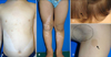
A 5-year-old boy had a white forelock and depigmented patches over the abdomen and extremities since birth. Physical examination revealed islands of pigmented macules within these leukodermic patches. He also had numerous café-au-lait macules on the trunk and extremities, at least six of which were > 5 mm in diameter (Fig. 1). The diagnosis of NF-1 is based on clinical criteria established by the National Institutes of Health Consensus Conference in 1987. According to these criteria, two or more of the following findings establish the diagnosis of NF-1; six or more café-au-lait macules > 5 mm in diameter in pre-pubertal individuals and > 15 mm in diameter after puberty; two or more neurofibromas or one plexiform neurofibroma; freckling in the axillary or inguinal regions; optic pathway tumor; distinctive bone lesions such as sphenoid wing dysplasia; and a first-degree relative with NF-13.
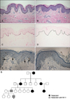
Our patient satisfied the diagnostic criteria. Skin biopsies were obtained from a depigmented lesion and a café-au-lait macule on the abdomen. Basal hyperpigmentation was noticed in the café-au-lait macule only. Fontana-Masson staining of the depigmented area was negative, while that of the café-au-lait macule revealed increased melanin pigments in the basal layer. No S-100-positive dendritic epidermal cells were detected in the leukodermic area, while some were detected in the pigmented macule (Fig. 2).
Family history revealed inheritance of piebaldism on the paternal side: the father was affected with a white forelock and several depigmented patches. In addition, the patient's mother referred to a paternal aunt and grandmother with similar depigmented patches (Fig. 2G). Among them, however, only his father had concomitant numerous café-au-lait macules. Based on the clinical and histological findings, we diagnosed this patient with piebaldism and NF-1.
To date, the co-occurrence of piebaldism and NF-1 has been described in only six patients including ours4,5. Piebaldism and NF-1 genes have been cloned and assigned to different chromosomes (4q12 and 17q11.2, respectively). Based on genetic and prevalence data, the co-occurrence of piebaldism and NF-1 appears most likely random. However, these two hereditary disorders are commonly related with abnormal melanocyte development and differentiation. By discovering additional genetic abnormalities and pathogeneses related to these disorders, we may be able to ensure early diagnosis, adequate follow-up, and genetic counseling for affected patients.
 XML Download