

 PDF
PDF ePub
ePub Citation
 Print
Print


Dear Editor:
Vitiligo is traditionally divided into two distinct clinical forms, nonsegmental vitiligo (NSV) and segmental vitiligo (SV). SV usually accompanies leukotrichia, which indicates that the condition is resistant to medical treatment and requires epidermal grafting1. In this case, perifollicular repigmentation was induced in a patient with SV and concurrent leukotrichia that was previously unresponsive to narrow-band ultraviolet B (NBUVB) phototherapy after the patient suffered an accidental burn on the denuded but uncovered area during phototherapy following an epidermal graft.
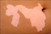
A 12-year-old girl presented to our clinic with white patches that had initially developed on the right side of her trunk 2 years prior. Physical examination showed well-defined depigmented patches containing white hairs along the right T6~T12 dermatomes (Fig. 1). White accentuation with obvious fluorescence was detected using Wood's lamp. She had received NBUVB phototherapy for 5 months without remarkable improvement. Upon consent, a suction blister epidermal graft was performed. Due to the sizeable lesion, several sessions of epidermal grafting were required.
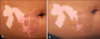
One week later, after we grossly concluded that the previously uncovered areas were fully recovered, phototherapy was initiated. After 3 days, the patient returned to the clinic only to display a burn on the formerly denuded but uncovered area. Further treatment was delayed until the area was completely healed, and 10 days later, treatment was re-engaged starting with 700 mJ/cm2, with an incremental 15% increase in dose during every subsequent course. Interestingly, after 2 weeks, spontaneous repigmentation at the denuded but uncovered site was observed along with that in the covered site (Fig. 2A). On the patient's subsequent visit 2 months later, the recipient area, again including the uncovered area, was almost completely pigmented (Fig. 2B).
Surgical treatment can be considered when vitiligo does not respond to traditional treatments. The procedure is particularly suitable in cases that show complete loss of melanocytes or in cases that have a low possibility of repigmentation that is clinically indicated by the presence of leukotrichia2. It has been suggested that visualization of leukotrichia in the vitiligous lesions of patients with SV using portable digital microscopy indicates no hope of repigmentation and, therefore, that a surgical approach is recommended1. A recent preliminary study, however, reported that although tyrosinase-positive melanocytes are only observed in black hair follicles, melanocytes still exist in the white hair follicles, although they are fewer in number than those in black hair follicles2. These findings suggest that the melanocyte reservoir is not completely depleted in vitiligo lesions showing leukotrichia.
According to an earlier study that revealed the promising result of combination treatment with an erbium:YAG laser and 5-fluorouracil ointment followed by NBUVB for the treatment of vitiligo, 78.1% of enrolled patients showed moderate to marked repigmentation3. In addition, microdermabrasion combined with pimecrolimus 1% cream has been reported to be effective without causing significant side effects including koebnerization in children with NSV4. The findings of these two studies suggest that epidermal damage caused by the erbium:YAG laser and microdermabrasion could induce the secretion of chemical mediators that are beneficial for melanocyte activation and enhance the NBUVB penetration and drug absorption into inactive melanocytes existing at the outer root sheath of the hair follicles.
Similarly, we believe that in our case, the sunburn of the lesion uncovered by a graft after phototherapy might have induced the formation of various chemical mediators and growth factors which could stimulate epidermal melanocytes and induce the proliferation and migration of inactive melanocytes in the outer root sheaths of hair follicles in the depigmented lesions5 despite the presence of leukotrichia. The burn itself would also increase the transmittance and accessibility of NBUVB into inactive melanocytes in the outer root sheath, further inducing perifollicular repigmentation.
In patients with SV, the coexistence of leukotrichia, has been considered a poor prognostic marker of disease that is often refractory to traditional treatment and eventually forces both the patient and the doctor into a surgical solution. Our case; however, shows that the few melanocytes that still remained in the hair follicles with leukotrichia could serve as a source of perifollicular repigmentation and points to an optimistic future involving treatment without the need for invasive epidermal grafting.
 XML Download