

 PDF
PDF ePub
ePub Citation
 Print
Print


Dear Editor:
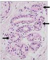
Argyria is a rare condition caused by improper exposure to products containing silver. Argyria will display as a blue-to-slate-gray color change in the field when exposed to sunlight1, and this may play an important role in the pigmentation. With respect to the pathologic findings, argyria is distinguished by the deposition of fine, brown-black granules mostly surrounding the basement membrane of the eccrine sweat glands and connective tissues in the dermis2. Unfortunately, an effective treatment is yet to be found. We experienced a example of argyria that showed noteworthy change after a Q-switched 1,064-nm Nd:YAG laser was used.
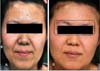
A 36-year-old woman was referred to the dermatologic department of Samsung Medical Center with a blue-gray color change of the face (Fig. 1A). According to her history, she had been ingesting colloidal silver for two years as a folk remedy. A dermatologic check up showed widespread blue-gray color change of the face. Histopathological examination results from her forehead skin showed spherical small brown-black granules deposited in the basement membrane around the eccrine glands, and scattered in the dermis (Fig. 2). The laboratory tests were unremarkable. The skin color change was dealt with using a Q-switched 1,064 nm Nd:YAG laser (Medlite IV; Continuum, Santa Clara, CA, USA). The laser was used at a fluence of 8 J/cm2 and pulse duration of 6 nanoseconds with a 4-mm spot size. There was no on-the-spot complications or any sequelae. Afterwards, we dealt with the patient's other part of the face after an interval of two to three months. Thereafter, with three lots of treatment, the treated area showed significant improvement (Fig. 1B). Agree on the use of patient photos obtained.
The pathophysiology of silver deposition in the development of argyria is not completely known. Like photography, sunlight reduces elemental silver to silver sul?de and selenide in the skin. This mechanism, combined with melanocyte stimulation, provokes cutaneous color changes3. Treatment of argyria is very difficult. The underlying mechanisms of argyria treatment by Q-switched lasers may be analogous to tattoo removal4. The 1,064-nm wave-length laser is weakly absorbed by melanin or water, which can penetrate into the deep dermis where eccrine sweat glands and silver granules exist. The present case showed significant improvement after four sessions of Q-switched Nd:YAG laser. The patient was highly satisfied with the result, and is continuously receiving the treatment to date. In conclusion, Q-switched 1,064-nm Nd:YAG laser is a useful modality for the treatment of argyria, even though more research is necessary to confirm whether the beneficial effect is coherent.
 XML Download