

 PDF
PDF ePub
ePub Citation
 Print
Print


Dear Editor:
Sialolithiasis is a common disease of the salivary glands. Most calculi occur in the major salivary glands such as the submandibular glands (80% to 92%) and parotid glands (16% to 19%), while minor salivary glands are rarely affected (2%)1. Minor salivary gland sialolithiasis is characterized by a small, solitary submucosal nodule, which is hard and in some cases can be movable in the surrounding tissue2. Since it is rare and its clinical features are not always typical, clinical misdiagnosis is possible3. Most otolaryngologists and dentists are relatively familiar with sialolithiasis, but many dermatologists are not. In order to heighten the awareness of this disease and to facilitate diagnosis, we report a case of minor salivary gland sialolithiasis that was initially misdiagnosed clinically.
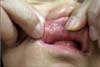
A 56-year-old man presented with a six-year history of an asymptomatic solitary submucosal nodule on the upper lip. He reported no history of trauma to the lip. Physical examination revealed a small (0.4×0.4 cm), firm, movable, well-defined submucosal nodule on the inner side of the upper lip (Fig. 1). We presumed the lesion was a mucocele and performed excisional biopsy. Histopathologic examination revealed heterogeneous lamellated calculi within the lumen of a dilated minor salivary duct. The calculi had a relatively homogenous core with alternating basophilic and eosinophilic bands at the periphery (Fig. 2A). The excretory ductal epithelium had undergone squamous metaplasia (Fig. 2B). These pathological features were consistent with the diagnosis of minor salivary gland sialolithiasis3,4.
A sialolith of the minor salivary glands is commonly described as a solitary, firm, asymptomatic, mobile nodule. It is most likely to develop near the upper lip and buccal mucosa that are susceptible to masticatory trauma4. Its clinical characteristics can resemble those of other lesions, resulting in frequent misdiagnosis. Anneroth et al. observed the correct diagnosis was made in only 20% of patients2. The differential diagnosis includes mucocele, foreign body, benign salivary neoplasm, and calcinosis cutis. A mucocele is clinically similar to minor salivary gland sialolithiasis. However, it can be readily diagnosed because of its cyst-like appearance lined by granulation tissues. A foreign body is difficult to diagnose unless imaged by radiography. It can only be confirmed after excision and microscopic examination. A benign minor salivary neoplasm tends to be an indolent, painless, firm, nodule, and the most frequent location is the same as minor salivary gland sialolithiasis. Microscopic examination is required to determine the exact histologic nature of the tumor. Several case reports have described calcinosis cutis of the lip, and this condition is difficult to differentiate from minor salivary gland sialolithiasis5. However, it reveals relatively homogeneous calcium deposits surrounded by epithelial-like cells, rather than heterogeneous lamellated calculi.
Sialolithiasis of the minor salivary glands is frequently misdiagnosed, mostly because its frequency, signs, and symptoms are relatively unknown, especially to dermatologists. We should be aware of this disease and consider it as a possible diagnosis when swelling of the oral mucosal tissues is observed in a patient.

 XML Download