

 PDF
PDF ePub
ePub Citation
 Print
Print


INTRODUCTION
Corals are coelenterates of the Anthozoa class1. They may cause injury either because of their nematocytes or due to their sharp-edged calcareous outer skeletons. Stings from their nematocytes may cause a rare type of contact dermatitis that can be divided into different reactions: acute, delayed and chronic coelenterate dermatitis. Acute reactions, such as acute urticaria and vesiculobullous lesions, can be due to the immediate effect of toxic substances contained in nematocytes, including quaternary ammonium compounds, proteins, 5-hydroxytryptamine, catecholamines, histamine and histamine liberators. Delayed reactions are less well known, but they are thought to be induced through both immediate and delayed hypersensitivity mechanisms. Chronic, persistent reactions are assumed to be a form of allergic contact dermatitis2.
We report here on a rare case of a delayed reaction of the skin to coral injury, and the patient displayed superficial granulomas. Among the different delayed reactions, granulomas have rarely been reported3. With the atypical CD30+ lymphocytes seen in this case, the delayed granulomatous reaction in our patient was considered to be an allergic foreign-body granuloma due to a hypersensitivity reaction and persistent antigen stimulation.
CASE REPORT
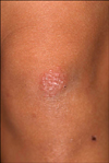
Several minutes after coming in contact with hard coral in the Gulf of Thailand, South China Sea, a 28-year-old female had a stinging, burning sensation on the resulting scratch wound on her left knee. Five days later, a pruritic erythematous patch composed of multiple flat papules developed at the site of the previous scratch (Fig. 1). No systemic symptoms were observed. The patient had no specific history of allergy. A biopsy was taken from the lesional skin. The epidermis showed acanthosis, spongiosis and focal lymphocyte exocytosis. A dense, superficial perivascular and periadnexal infiltrate of lymphocytes and eosinophils was present in the dermis. Focal superficial small epithelioid granuloma with multinucleated giant cells and focal collagen degeneration was also present (Fig. 2A). Observation under polarized light failed to demonstrate any foreign material inside the granulomas. On the immunohistochemistry, CD3+ and CD4+ T-helper (Th) cells, S-100+ dendritic cells and CD30+ nucleolated atypical lymphocytes were revealed in the inflammatory infiltrates (Fig. 2B, C). After one month of topical corticosteroid application, the skin lesions cleared completely with leaving some residual hypopigmentation.
DISCUSSION
Coral dermatitis can be divided into three or four stages. Miracco et al2 divided it into the acute, delayed and chronic stages and Addy4 classified it as acute urticaria, acute vesiculobullous dermatitis, subacute fleshy granulomatous dermatitis and chronic lichenoid dermatitis. These stages does not have strict definitions, but the clinically acute stage usually develops immediately or within several hours after contact with coral and it resolves within several hours or days. The delayed stage can develop several days or weeks after this. The chronic stages can be recurrent or the lichenoid dermatitis can persist. The histology of the acute stages show spongiosis with microvesicles, intracellular edema of the epidermis and edema of the papillary dermis. The features of the delayed stages are similar to those of pityriasis lichenoides and persistent insect-bite reaction, showing wedge shaped, subepidermal and perivascular mononuclear cell infiltrates and extravasated erythrocytes. The chronic stages show acanthosis, parakeratosis and an increased granular layer, in addition to a diffuse dense mononuclear cell infiltration in the upper dermis2,4.
The skin lesions in the current case are clinically and pathologically similar to those described in the cases of delayed skin reaction to coelenterates, except this case also showed superficial granulomas. Miracco et al2 reported a case that displayed a delayed skin reaction to coral injury with superficial granulomas and atypical CD30+ lymphocytes. The same results were obtained on immunohistochemical staining. We observed a predominance of Langerhans cells and Th lymphocytes in the inflammatory infiltrates, as well as focal epithelioid granulomas and numerous large CD30+ nucleolated lymphocytes.
It has generally been assumed that most coelenterate envenomation reactions are toxic rather than allergic, but there have been several reports of delayed, recurrent, persistent skin reactions that have been attributed to allergies. In additional to type I hypersensitivity, type IV allergy is considered to be involved in some such eruptions5. Addy et al4 showed coral dermatitis to be an allergic contact dermatitis because not all people who have contact with coral actually develop the dermatitis. That study suggested some of the risk factors for coral dermatitis are seafood allergy and atopic dermatitis.
The granuloma of coral dermatitis is considered to be a foreign-body reaction. Generally, the foreign-body granulomas can be divided into two groups: nonallergic and allergic. Nonallergic foreign-body reaction typically shows a granulomatous response that's marked by histiocytes and giant cells surrounding the foreign material. Often, some of the giant cells are of the foreign-body type. Polariscopy is helpful for identification of foreign materials. Allergic foreign-body granuloma shows a sarcoidal or tuberculoid pattern that consists of epithelioid cells with or without giant cells. Phagocytosis of the foreign substance is very slight or absent. Some substances that first act as foreign material may later on act as allergens after sensitization has occurred, as in the case of seaurchin spines and silica6. In this case, the superficial granuloma was considered to be caused by allergic reaction. The clinically delayed onset of the skin lesions, the pathologically epithelioid cell infiltrations with Langerhans giant cells and no foreign material found by polariscopy suggest allergic reaction was responsible for the patient's condition.
The CD30 (Ki-1) antigen expression on T cells has been proposed to be a marker for a subset of memory T cells with potent B cell helper activity7. Atypical CD30+ T cells are frequently observed in lymphoproliferative disorders and acute atopic dermatitis, scabies and EBV, HIV, HTLV and hepatitis B&C virus infection. The presence of atypical CD30+ lymphocytes can be considered to be due to hypersensitivity reaction and persistent antigen stimulation. The described case is the second case report showing superficial granuloma formation with CD30+ T cells in a patient with delayed coral dermatitis. The case of Miracco et al2 is quite similar to our case, except their patient suffered from atopic dermatitis. Because CD30+ lymphocytes are frequently found in atopic patients8, they may not be associated with delayed coral dermatitis. However, because our case did not have a history of atopic dermatitis, we can suggest there was a more reliable association between CD30+ lymphocytes and the delayed granulomatous coral dermatitis. Further studies are needed to determine the function of the CD30+ lymphocytes in delayed coral dermatitis, and especially in the resulting granuloma.

 XML Download