

 PDF
PDF ePub
ePub Citation
 Print
Print


Abstract
Cerebrospinal fluid (CSF) rhinorrhea is classified into traumatic and non-traumatic types. Traumatic CSF rhinorrhea comprises the majority of cases, and major causes include head trauma, rhinologic procedures, and neurosurgery. Non-traumatic (spontaneous) CSF rhinorrhea with normal cerebrospinal pressure is a rare condition, occurring in only 4% of cases. We recently experienced a case of spontaneous CSF rhinorrhea complicated with bacterial meningitis. The defect site was identified in the left sphenoid sinus and was successfully repaired with a nasoseptal flap under an endoscopic approach. We present the etiology, classification, and treatment of this rare disease entity with a review of the literature.
REFERENCES
1). Citardi MJ, Fakhri S. Cerebrospinal fluid rhinorrhea. Flint PW, Haughey BH, Lund V, Niparko JK, Robbins KT, Thomas JR, editors. Cummings Otolaryngology-Head and Neck Surgery. 6th ed.Philadelphia, PA: Elsevier Saunders;2015. p. 803–15.

2). Loew F, Pertuiset B, Chaumier EE, Jaksche H. Traumatic spontaneous and postoperative cerebrospinal fluid rhinorrhea. Adv Tech Stand Neurosurg. 1984; 11:169–207.
3). Shugar JM, Som PH, Eisman W, Biller HF. Non-traumatic cerebrospinal fluid rhinorrhea. Laryngoscope. 1981; 91(1):114–20.
4). Daudia A, Biswas D, Jones NS. Risk of meningitis with cerebrospinal fluid rhinorrhea. Ann Otol Rhinol Laryngol. 2007; 116(12):902–5.
5). Katz RT, Kaplan PE. Glucose oxidase sticks and cerebrospinal fluid rhinorrhea. Arch Phys Med Rehabil. 1985; 66(6):391–3.
6). berascher G, Arrer E. Efficiency of various methods of identifying cerebrospinal fluid in oto- and rhinorrhea. ORL J Otorhinolaryngol Relat Spec. 1986; 48(6):320–5.
7). Lloyd KM, DelGaudio JM, Hudgins PA. Imaging of skull base cerebrospinal fluid leaks in adults. Radiology. 2008; 248(3):725–36.
8). Ziu M, Savage JG, Jimenez DF. Diagnosis and treatment of cerebrospinal fluid rhinorrhea following accidental traumatic anterior skull base fractures. Neurosurg Focus. 2012; 32(6):E3.
9). Hegazy HM, Carrau RL, Snyderman CH, Kassam A, Zweig J. Trans-nasal endoscopic repair of cerebrospinal fluid rhinorrhea: a meta-analysis. Laryngoscope. 2000; 110(7):1166–72.
10). Martin TJ, Loehrl TA. Endoscopic CSF leak repair. Curr Opin Otolaryngol Head Neck Surg. 2007; 15(1):35–9.
11). Gassner HG, Ponikau JU, Sherris DA, Kern EB. CSF rhinorrhea: 95 consecutive surgical cases with long term follow-up at the Mayo Clinic. Am J Rhinol. 1999; 13(6):439–47.
12). Hadad G, Bassagasteqguy L, Carrau RL, Mataza JC, Kassam AB, Snyderman CH, et al. A novel reconstructive technique after endoscopic expanded endonasal approaches: vascular pedicle nasoseptal flap. Laryngoscope. 2006; 116(10):1882–6.
13). Lee KH, Yang CW. Endoscopic endonasal skull base repair with nasoseptal flap. Korean J Otolaryngol. 2015; 58(1):7–11.
14). Lopatin AS, Kapitanov DN, Potapov AA. Endonasal endoscopic repair of spontaneous cerebrospinal fluid leaks. Arch Otolaryngol Head Neck Surg. 2003; 129(8):859–63.
15). Schlosser RJ, Bolger WE. Nasal cerebrospinal fluid leaks: critical review and surgical considerations. Laryngoscope. 2004; 114(2):255–65.
Fig. 1.
Axial ( A) and coronal ( B) sections of preoperative computed tomography and magnetic resonance imaging ( C). The arrows indicate fluid collection in the left sphenoid sinus.
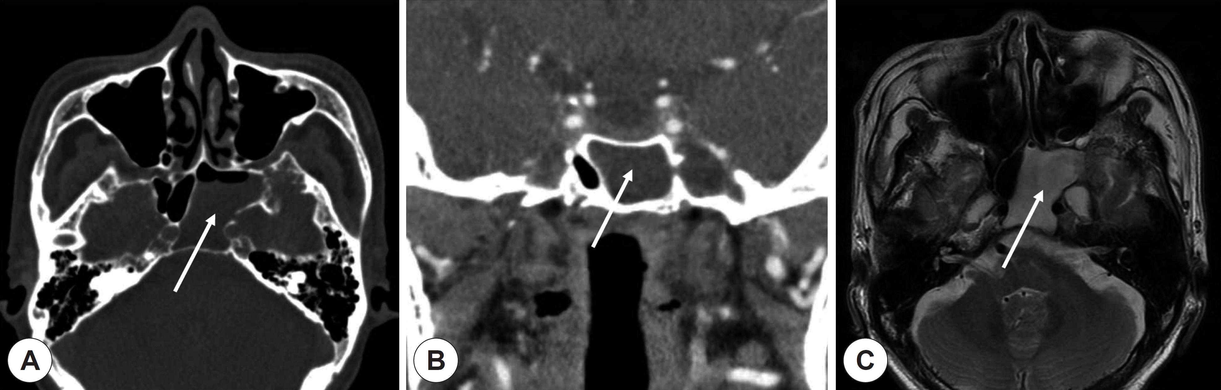
Fig. 2.
Findings of metrizamide computed tomography cister-nography. Arrow indicates bony defect of posterolateral wall of the left sphenoid sinus.
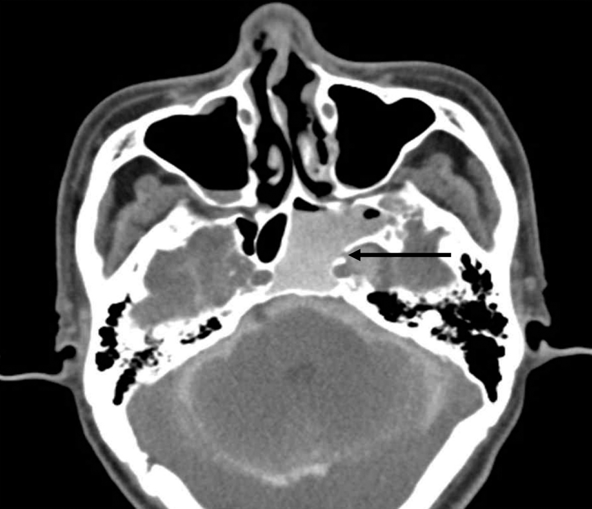
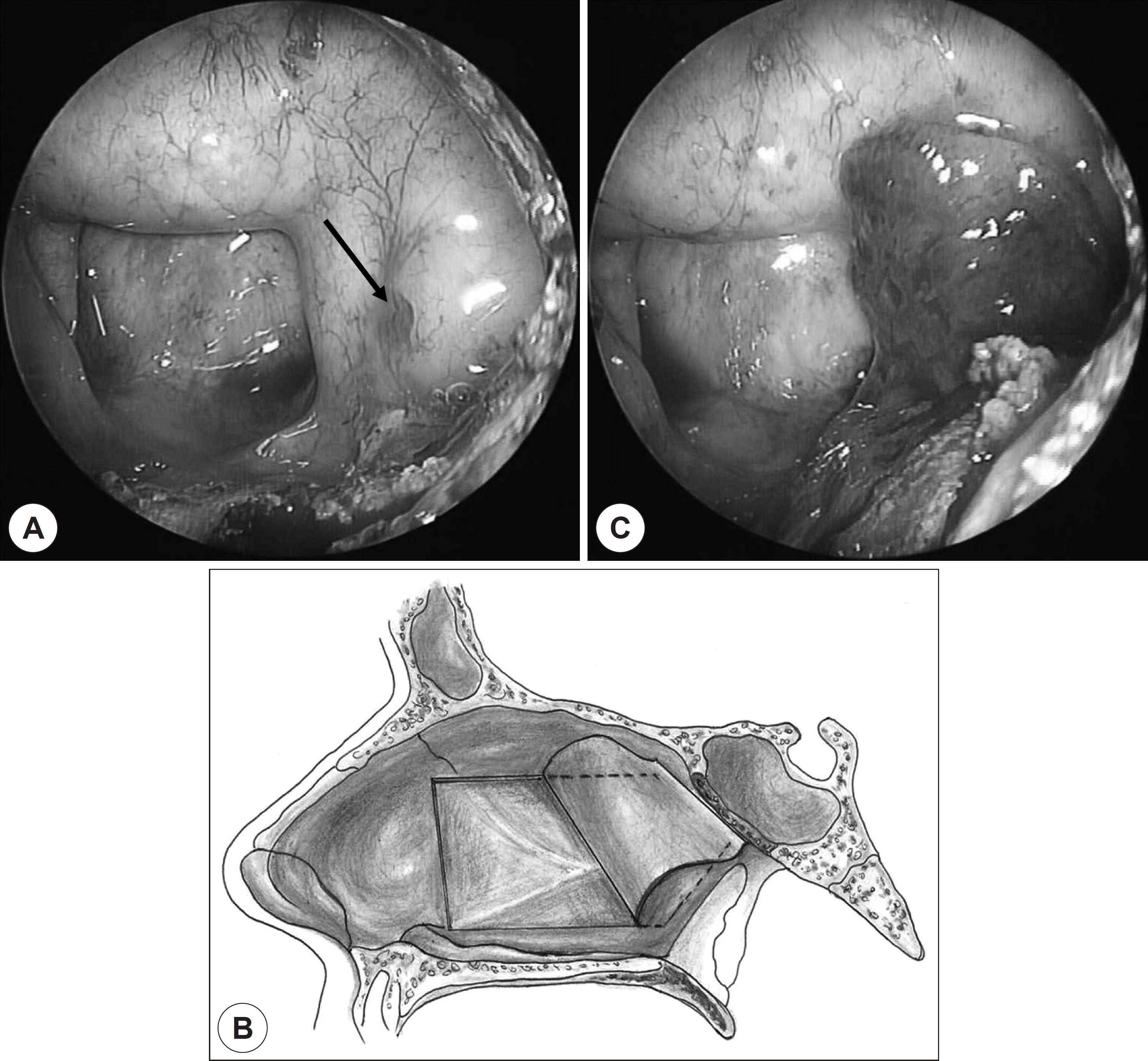
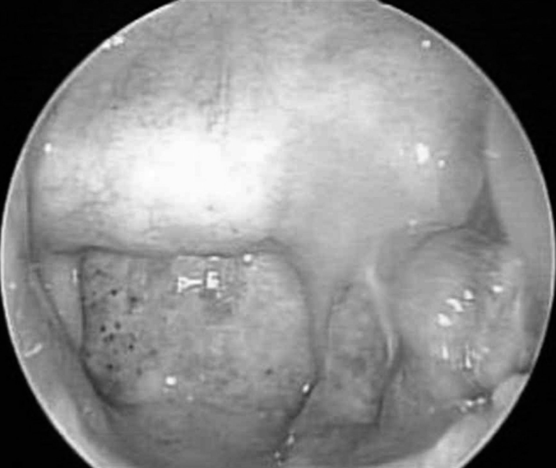
Fig. 3.
Endoscopic findings and schematic illustration of pedicled nasoseptal flap. Cerebrospinal fluid leakage was identified at the posterolateral wall of the left sphenoid sinus ( arrow) ( A). Harvest of the nasoseptal flap ( B). Cerebrospinal fluid leakage site was cov-ered with nasoseptal flap ( C).
 XML Download