Abstract
Schwannomas are benign neoplasms arising from the sheath of myelinated nerve fibers and may occur in any part of the body. They mostly occur in the head and neck region, accounting for about 25% to 45% of all cases. The eighth cranial nerve is the most common site of origin. About 4% of all head and neck schwannomas originate in the nasal cavity and paranasal sinuses. The best treatment of schwannomas is surgical excision. Since it is an encapsulated tumor, difficultly is rarely encountered in its complete removal, and recurrence is unlikely. We present a unique and rare case of a 71-year-old man with a recurrent septal mass, finally diagnosed as a schwannoma, with a review of the literature.
References
1). Jung SG, Han SY, Kim DE, Ahn BH. A case of schwannoma of the nasal septum. Korean J Otorhinolaryngol-Head Neck Surg. 2010; 53:497–500.

2). Berlucchi M, Piazza C, Blanzuoli L, Battaglia G, Nicolai P. Schwannoma of the nasal septum: a case report with review of the literature. Eur Arch Otorhinolaryngol. 2000; 257(7):402–5.

4). Pasquini E, Sciarretta V, Farneti G, Ippolito A, Mazzatenta D, Frank G. Endoscopic endonasal approach for the treatment of benign schwannoa of the sinonasal tract and pterygopalatine fossa. Am J Rhinol. 2002; 16(2):113–8.
5). Srinivasan V, Deans JA, Nicol A. Sphenoid sinujs schwannoma treated by endoscopic excision. J Laryngol Otol. 1999; 113(5):466–8.
6). Seo YI, Nam SY, An KH, Kim SY, Lee KS. Extracranial nerve sheath tumors of the head and neck. Korean J Otorhinolaryngol-Head Neck Surg. 1997; 40(6):908–13.
7). Park EH, Lee SS, Byun SW. A schwannoma in the nasal septum. Eur Arch Otorhinolaryngol. 2008; 265:983–5.

8). Wang LF, Tai CF, Chai CY, Ho KY, Kuo WR. Schwannoma of the nasal septum: a case report. Kaohsiumg J Med Sci. 2004; 20(3):142–5.

9). Koh TK, Bae WY, Rha SH. A case of nasal schwannoma coexisting with epidermal cyst. Korean J Otorhinolaryngol-Head Neck Surg. 2013; 56:787–90.

10). Zhou P, Zeng F, Li J, Liu S. Only septal deviation? A tiny schwannoma in the nasal septum. Indian J Otolaryngol Head Neck Surg. 2013; 65(1):80–2.

11). Rajagopal S, Kaushik V, Iron K, Herd ME, Bhatnagar RK. Schwannoma of the nasal septum. Br J Radiol. 2006; 79(943):e16–8.

12). Wada A, Matsuda H, Matsuoka K, Kawano T, Furukawa S, Tsukuda M. A case of schwannoma on the nasal septum. Auris Nasus Larynx. 2001; 28(2):173–5.

13). Choe H, Jun YJ, Cho WS, Kim TH. A case of schwannoma of the nasal cavity mimicking olfactory neuroblastoma. Korean J Otolaryngol Head Neck Surg. 2007; 50:548–51.
14). Tsai PY, Chan MY, Chen SH, Lin CY, Chen PH. The prognosis and recurrence of head and neck schwannomas: An 8-year retrospective study. Taiwan J Oral Maxillofac Surg. 2011; 22:165–74.
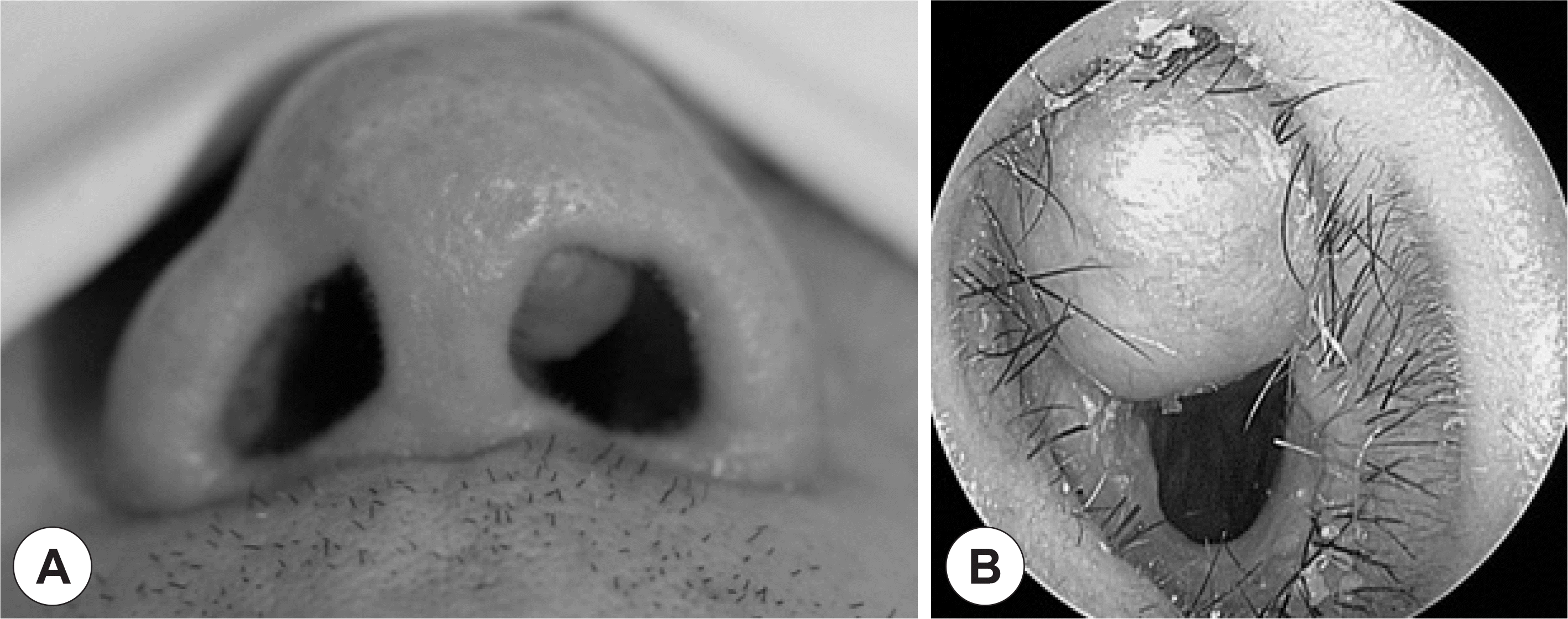
Fig. 1.
These photos show about 1.5×1.2 cm sized mass on left side of nasal septum. A: Basal view. B: Endoscopic finding.

Fig. 2.
The PNS CT and Brain MRI findings. These show about 1.5×1.2 cm sized non-enhanced mass in caudal septum. A: Enhanced axial view. B: Enhanced coronal view. C: The mass shows intermediate signal intensity in T1 weighted image. D: T2 Fat-saturation image shows the mass with high signal intensity.

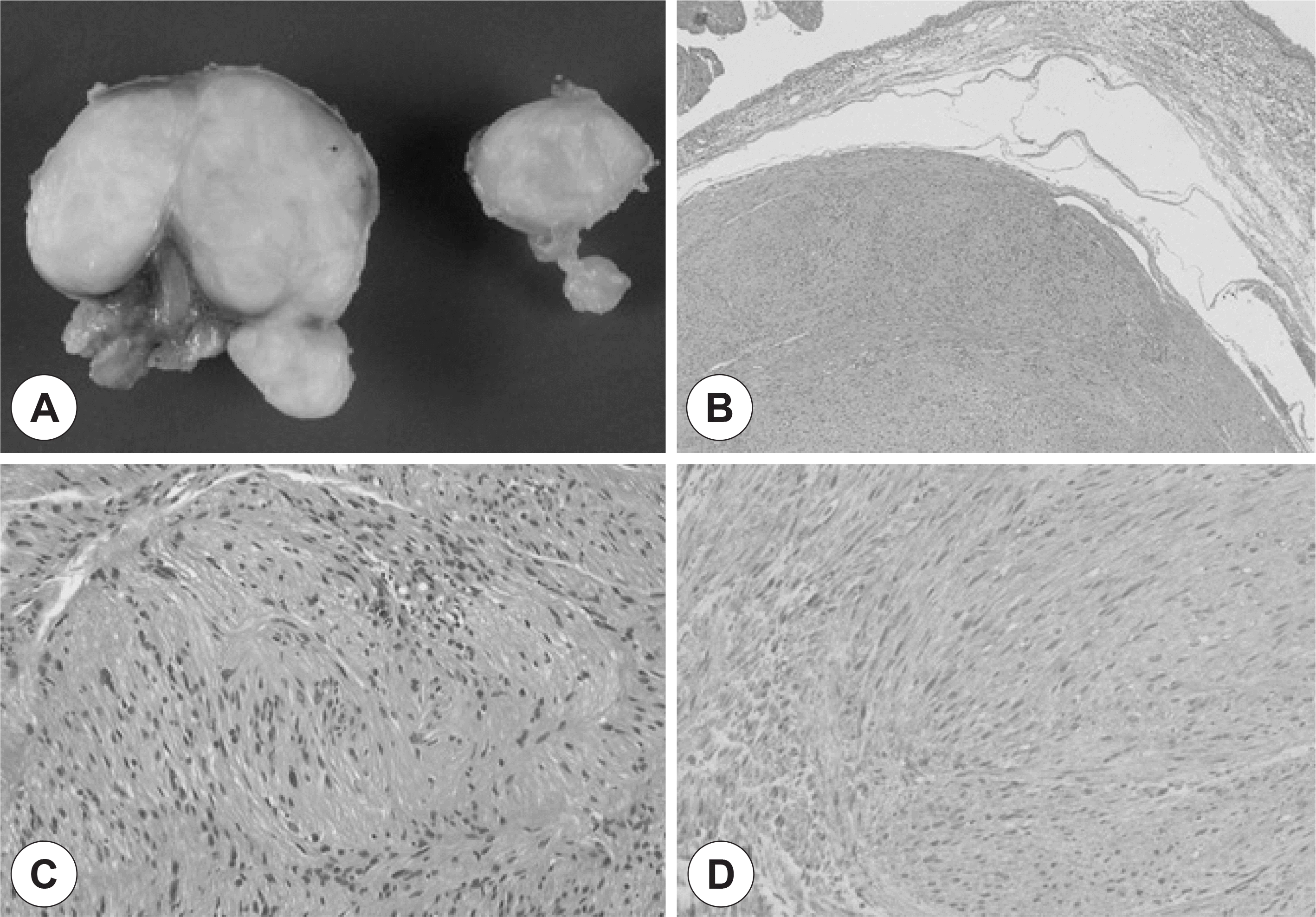
Fig. 3.
Pathologic findings. A: Gross photo shows two well-circumscribed masses. One is about 1.8×1.3 cm sized and the other is 1.0×0.8 cm sized. B: The tumor is well circumscribed (H&E, ×40). C: This shows characteristic nuclear palisading (Verocay bodies) (H&E, ×200). D: The tumor cells are immunoreactive for S-100 protein (S-100, ×200).


 PDF
PDF ePub
ePub Citation
 Print
Print


 XML Download