

 PDF
PDF ePub
ePub Citation
 Print
Print


Abstract
Objective
To investigate whether the accuracy of 3D laser scanning is influenced by the angles and number of scans.
Methods
Using a 3D laser scanner, 10 manikins with facial markers were scanned at 7 horizontal angles (front view and at 20°, 45°, and 60° angles on the right and left sides). Three-dimensional facial images were reconstructed by 6 methods differing in the number and angles of scans, and measurements of these images were compared to the physical measurements from the manikins.
Results
The laser scan images were magnified by 0.14 - 0.26%. For images reconstructed by merging 2 scans, excluding the front view; and by merging 3 scans, including the front view and scans obtained at 20° on both sides; several measurements were significantly different than the physical measurements. However, for images reconstructed by merging 3 scans, including the front view; and 5 scans, including the front view and scans obtained at 20° and 60° on both sides; only 1 measurement was significantly different.
Figures and Tables
Fig. 1
The positions of the facial markers used in this study: FRt, right area of forehead; FLt, left area of forehead; Fm, middle area of forehead; G, glabella; N', soft tissue nasion; Pn, pronasale; ExRt, right exocanthion; ExLt, left exocanthion; ZyRt, right zygion; ZyLt, left zygion; CkRt, right cheek; CkLt, left cheek; ChRt, right cheilion; ChLt, left cheilion; UL, upper lip; LL, lower lip; Go'Rt, right soft tissue gonion; Go'Lt, left soft tissue gonion; Pg', soft tissue pogonion; TraRt, right tragus; TraLt, left tragus.
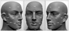
Fig. 2
The laser scanner and rotator used in this study. A, Vivid 910 3D laser scanner (Minolta, Tokyo, Japan); B, rotator fabricated for this study. Protractor is attached at the base of the rotator.
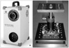
Fig. 3
Linear measurements used in this study: 1, Pn-Fm; 2, Pn-FRt; 3, Pn-FLt; 4, Pn-G; 5, Pn-N'; 6, Pn-ExRt; 7, Pn-ExLt; 8, Pn-ZyRt; 9, Pn-ZyLt; 10, Pn-CkRt; 11, Pn-CkLt; 12, Pn-ChRt; 13, Pn-ChLt; 14, Pn-UL; 15, Pn-LL; 16, Pn-Pg'; 17, Pn-Go'Rt; 18, Pn-Go'Lt; 19, Pn-TraRt; 20, Pn-TraLt; 21, FRt-FLt; 22, ExRt-ExLt; 23, ZyRt-ZyLt; 24, CkRt-CkLt; 25, ChRt-ChLt; 26, Go'Rt-Go'Lt
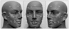
Fig. 4
Integrated 3D laser scan images were created in RapidForm software, and measurements were obtained using the 3D measure function.
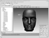
Fig. 5
Bland-Altman plots for variations in (Pn-Pg') of Methods 4, 5, and 6. The x-axis shows the average of each integrated 3D laser scan image with physical measurements of manikins, whereas the y-axis represents the difference between the 2 measurements. The limits of agreement are indicated by horizontal lines.
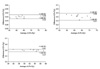
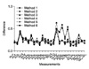
Fig. 6
Differences between each integrated 3D laser scan image and the physical measurements of the manikins (mm).
References
1. Mah J, Hatcher D. Current status and future needs in craniofacial imaging. Orthod Craniofac Res. 2003. 6:Suppl 1. 10–16.

2. Kusnoto B, Evans CA. Reliability of a 3D surface laser scanner for orthodontic applications. Am J Orthod Dentofacial Orthop. 2002. 122:342–348.
3. Kau CH, Richmond S, Zhurov AI, Knox J, Chestnutt I, Hartles F, et al. Reliability of measuring facial morphology with a 3-dimensional laser scanning system. Am J Orthod Dentofacial Orthop. 2005. 128:424–430.
4. Kau CH, Richmond S, Savio C, Mallorie C. Measuring adult facial morphology in three dimensions. Angle Orthod. 2006. 76:773–778.
5. Baik HS, Lee HJ, Jeon JM. A proposal of soft tissue landmarks for craniofacial analysis using three-dimensional laser scan imaging. Korean J Orthod. 2006. 36:1–13.
6. Kau CH, Zhurov A, Richmond S, Cronin A, Savio C, Mallorie C. Facial templates: a new perspective in three dimensions. Orthod Craniofac Res. 2006. 9:10–17.
7. Baik HS, Jeon JM, Lee HJ. A study of facial soft tissue of Korean adults with normal occlusion using a three-dimensional laser scanner. Korean J Orthod. 2006. 36:14–29.
8. Kau CH, Zhurov A, Richmond S, Bibb R, Sugar A, Knox J, et al. The 3-dimensional construction of the average 11-year-old child face: a clinical evaluation and application. J Oral Maxillofac Surg. 2006. 64:1086–1092.
9. Moss JP, Linney AD, Lowey MN. The use of three-dimensional techniques in facial esthetics. Semin Orthod. 1995. 1:94–104.
10. Kau CH, Zhurov A, Bibb R, Hunter L, Richmond S. The investigation of the changing facial appearance of identical twins employing a three-dimensional laser imaging system. Orthod Craniofac Res. 2005. 8:85–90.
11. Hennessy RJ, Moss JP. Facial growth: separating shape from size. Eur J Orthod. 2001. 23:275–285.
12. McDonagh S, Moss JP, Goodwin P, Lee RT. A prospective optical surface scanning and cephalometric assessment of the effect of functional appliances on the soft tissues. Eur J Orthod. 2001. 23:115–126.
13. Ismail SF, Moss JP, Hennessy R. Three-dimensional assessment of the effects of extraction and nonextraction orthodontic treatment on the face. Am J Orthod Dentofacial Orthop. 2002. 121:244–256.
14. Moss JP, Ismail SF, Hennessy RJ. Three-dimensional assessment of treatment outcomes on the face. Orthod Craniofac Res. 2003. 6:Suppl 1. 126–131.
15. Kau CH, Cronin A, Durning P, Zhurov AI, Sandham A, Richmond S. A new method for the 3D measurement of postoperative swelling following orthognathic surgery. Orthod Craniofac Res. 2006. 9:31–37.
16. McCance AM, Moss JP, Fright WR, Linney AD, James DR. Three-dimensional analysis techniques--Part 2: Laser scanning: a quantitative three-dimensional soft-tissue analysis using a color-coding system. Cleft Palate Craniofac J. 1997. 34:46–51.
17. Ferrario VF, Sforza C, Poggio CE, Serrao G. Facial three-dimensional morphometry. Am J Orthod Dentofacial Orthop. 1996. 109:86–93.
18. Choi HH, Cho JH, Park HJ, Oh HK, Choi JH, Hwang HS, et al. Validity of superimposition range at 3-dimensional facial images. J Korean Assoc Maxillofac Plast Reconstr Surg. 2009. 31:149–157.
19. Broadbent BH. A new x-ray technique and its application to orthodontia. Angle Orthod. 1931. 1:45–66.
20. Sanborn RT. Differences between the facial skeletal patterns of Class III malocclusion and normal occlusion. Angle Orthod. 1955. 25:208–222.
21. Steiner CC. The use of cephalometrics as an aid to planning and assessing orthodontic treatment. Am J Orthod. 1960. 46:721–735.
22. Song JM, Lee KH, Hwang HS. A comparative study of guiding methods for natural head posture in cephalometrics. Korean J Orthod. 2005. 35:341–350.
23. Kim EH, Hwang HS. The validity of head posture aligner in posteroanterior cephaiometry. Korean J Orthod. 2000. 30:543–552.
24. Downs WB. Analysis of the dentofacial profile. Angle Orthod. 1956. 26:191–212.
25. Besl PJ, Mckay ND. A method for registration of 3-D shapes. IEEE Trans Pattern Anal Mach Intell. 1992. 14:239–256.
 XML Download