

 PDF
PDF ePub
ePub Citation
 Print
Print


INTRODUCTION
Manufacturers have continuously introduced new adhesives in dentistry that are more reliable, i.e. stronger, adhere better, less prone to leakage at margins and/or easier to handle.1 As new materials and techniques are introduced, orthodontists adopt some of these innovations and add them to their armamentarium,1 including the use of self-etching primers, resin modified glass ionomer cement (RMGIC), chlorhexidine varnishes and different adhesives etc.
Odontogenesis, a series of events taking place from bud formation stage until the completion of calcification and maturation of the tooth, is a complex process.2 Upon eruption, the outermost layer of enamel is immature and not fully calcified.2 This outer layer then begins to calcify due to the effects of salivary minerals. Although not well understood, it is known that changes in both mineral and organic components of the enamel are involved during post-eruptive maturation.3 It has been shown that the hydroxyl (OH-) group of hydroxyl-apatite crystals is absent in less mature enamel, but it can be found in mature enamel by using Fourier-transform-infrared-spectrometry.4 Ooya5 carried out a scanning electron microscope (SEM) study and showed that lingual and buccal surfaces of mature teeth have a prism-less enamel structure. However, the same areas of newly erupted teeth are prismatic.5 Compositional analyses of successive layers of enamel suggest that mineralization can take place to a depth of 0.5 mm for some time after eruption.6 Since there are structural differences between mature and newly erupted teeth, it is logical to expect differences in bond strengths of orthodontic attachments bonded to mature teeth versus newly erupted or unerupted teeth.2
The acid-etch bonding technique is commonly used in orthodontic clinics for attaching brackets. For bonding application, phosphoric acid etching is recommended for composite resin adhesives and poly-acrylic acid etching for resin-modified glass-ionomer cements (RMGIC),7 however both of these etching techniques require rinsing and air-drying. To simplify orthodontic bonding, self-etching primer (SEP) systems, which combines the steps of acid etching, rinsing and priming8 reduces the clinical steps and saves clinical operation time, because the procedure requires simply air-drying after application. According to White9 SEPs are easily manipulated and used, resulting in comfort for the patients and decreasing the chair time by 65%.
Because newly erupted or unerupted teeth have compositional and structural differences in their enamel minerals,6,10 less etching time might be needed to create the surface irregularities required for bonding, or more time might be needed for etching mature teeth.11
Tüfekçi et al.11 investigated the differences in shear bond strength (SBS) between newly erupted (taken from 13 - 14 year old patients) and mature (taken from >23 year old patients) premolar teeth when using both conventional and self-etching techniques for bonding orthodontic appliances and concluded that bond strength does not appear to be affected by the post-eruptive enamel maturation process. Jacobs et al.12 investigated the acid etching times and bonding characteristics of erupted and impacted teeth from young (12 to 24 years of age) and older (over 50 years of age) persons. They found that, differences in composition and surface structure of enamel between unerupted teeth and those that had been exposed to the oral environment do not appear to be large enough to cause a statistically significant difference in bond strength. Oliver13 evaluated the SBS of orthodontic attachments to enamel by using conventional adhesive systems, from unerupted and erupted young permanent teeth and their results gave no significant difference in bond strength between the two groups.
No research has been published in the literature that has compared the SBS values and failure-modes of orthodontic attachments bonded to unerupted teeth with conventional and self-etching adhesive systems.
Thus, the aim of this study was to evaluate and compare the SBS and adhesive remnant index (ARI) scores of orthodontic buttons bonded with conventional and self-etching adhesives to erupted and unerupted teeth. For the purposes of this study, the null hypothesis assumed that there were no statistically significant differences between the SBS values and the site of bond-failure of orthodontic buttons bonded to erupted and unerupted teeth that prepared by conventional and self-etching methods.
MATERIAL AND METHODS
One hundred sixty-eight extracted, sound, human third-molar teeth were used in the study. The criteria for tooth selection included: intact enamel not subjected to any pretreatment chemical agents (e.g. hydrogen peroxide), no cracks and gross-irregularities and no caries. Two groups of specimens were equally prepared according to the developmental stage of the teeth: erupted and unerupted. Teeth were collected from patients between ages of 18 and 30 years. The "erupted teeth" were completely erupted into the oral cavity with no surfaces covered by gingival soft tissue. The "unerupted (impacted) teeth" were those teeth that had no exposure to the oral cavity; they included both soft- and hard-tissue impactions. Following extraction, the teeth were immediately placed in distilled water at room-temperature and stored until the bonding procedure. The root of each tooth was embedded into an acrylic (Imicryl, Konya, Turkey) cylindrical block.
Metallic buttons (G&H Wire, Greenwood, USA) were used in the study. The average button base surface area was determined to be 9.43 mm2 from the manufacturer's instructions.
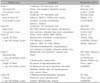
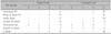
Table 1 shows the primer and adhesive systems that were used in the current study. The buttons were bonded to the mounted teeth following one of theseven adhesive protocols according to the manufacturers' instructions. Each group contained 12 specimens. Conventional etching and adhesive systems were used in Groups I - III; and self-etching systems in groups IV - VII.
A 37% phosphoric acid gel (Ventura Gel Acondicionador, Madespa, Spain) was applied to the enamel for 15-seconds and the teeth were then rinsed with water spray for 30-seconds and air dried for 20 seconds. After surface preparation, liquid primer was applied to the etched surface in the conventional groups.
Activation procedures for the self-etching primer were performed according to the manufacturer's instructions. Self-etching primer (Table 1) was gently rubbed onto the enamel surface for approximately three seconds with the disposable applicator supplied with the system. Then, a moisture-free air source was used to deliver a gentle burst of air to the enamel.
After etching/priming, bonding agent was photopolymerized in all groups for 10-seconds. To exclude possible differences in bond strength caused by the orthodontic composite used, the same material (Transbond XT, 3M Unitek, Monrovia, CA, USA) was applied under all buttons. Standard edgewise premolar stainless-steel brackets (3M Unitek, Monrovia, CA, USA) were positioned in the center of the crown and firm pressure was applied. Any excess composite was removed. Before light-curing, the buttons were slightly pressed with bracket holder and excess adhesive was removed with a scaler. A light-emitting diode (LED) (SmartLite, Dentsply, Milford, USA) was used for curing the composite, 20 seconds from both the mesial and distal sides. The same clinician carried out all bonding procedures in all groups (C.Y.). The teeth were then placed in distilled water at 37℃ for 24 hours before testing.
Debonding procedure
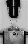
Each toothwas oriented with a guiding device, so that its tooth surfaces were parallel to the shear-force during the test. A gingivo-occlusal load was applied to the button, producing a shear force from the button (Fig 1). A computer, electronically connected to the Lloyd testing machine (Lloyd instruments, Foreham, Hampshire, UK), recorded the results of each test. The SBSs were measured at a crosshead speed of 1 mm/min. The force required to remove the buttons was measured in Newtons (N), and the SBS (1 megapascal-MPa = 1 N/mm2) was then calculated by dividing the force values by the button base area (9.43 mm2).
Evaluation of the residual adhesive
After debonding, all the teeth and buttons were evaluated under a stereomicroscope (Nikon, SMZ-1B, Osaka, Japan) by another operator (M.B.) who was blinded to the group allocation, under 10× magnification for the adhesive remnant index (ARI)14 scores: 0, no adhesive remaining on tooth; 1, less than half of the enamel bonding site covered with adhesive; 2, more than half of the enamel bonding site covered with adhesive; 3, the enamel bonding site covered entirely with adhesive.
Scanning electron microscope evaluation
For SEM investigations erupted and unerupted tooth specimens were used to evaluate the enamel surfaces. Tooth were transferred to 70% ethanol and dehydrated in increasing concentrations of ethanol. Specimens were gradually dehydrated through a graded series of ethanol, air-dried and mounted on SEM stubs so that the relevant area of interest could be seen, sputter coated with 10 nm of platinum in a Polaron E5100 SEM coating unit (Polaron Equipment Ltd, Hertfordshire, England), and examined in a Hitachi S 2500 SEM (Hitachi Ltd, Tokyo, Japan) at an operating voltage of 15 kV. The SEM photomicrographs were taken at 500× and 1500× magnification for visual inspection.
Statistical analysis
The Shapiro-Wilks normality test and the Levene variance homogeneity test were applied to the SBS data. The data showed normal distribution, and there was homogeneity of variances between the groups. Thus, the statistical evaluation of SBS values between test groups was performed using parametric tests.
Descriptive statistics, including mean, standard deviation, minimum and maximum values were calculated for all groups of the erupted and unerupted teeth. An independent sample t-test was undertaken to compare the SBS values of the same adhesive system between the erupted and unerupted teeth groups. The SBS values were analyzed by one-way analysis of variance (ANOVA) to determine significance of differences among 7 adhesive systems for each tooth type (erupted and unerupted), separately. To analyze the failure sites, contingency tables were designed and subjected to the chi-square test. The statistical significance level was established at p < 0.05.
Scoring of the ARI scores were repeated 4 months after the first measurement. Paired sample t-test was applied to the first and second data. It was found that the differences between the first and second measurements of the ARI scores were insignificant. The intra-observer intraclass correlation coefficient was 0.90 for erupted teeth and 0.94 for unerupted teeth.
RESULTS
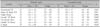
The descriptive statistics and the results of independent sample t-test are presented in Table 2. When the SBS values of erupted and unerupted teeth were compared, significant differences were found in two self-etching adhesives (Clearfil SE Bond and G Bond). Bond strengths of all adhesive systems were higher in unerupted teeth than erupted teeth, except for the Single-Bond system. Thus, the SBS part of the null hypothesis of this study was rejected.
The ANOVA comparisons of the 7 bonding systems for erupted and unerupted teeth groups are shown in Table 3 and Table 4, respectively. No statistically significant differences were found among 7 adhesive groups for both erupted and unerupted teeth (p > 0.05).
The ARI scores for the adhesive systems are listed in Table 5. The data distributions indicated that bond failure occurred more frequently at the button-adhesive interface. For both teeth groups and adhesive systems the distribution of the ARI scores was similar and showed no significant differences. Thus, the failure-mode part of the present null hypothesis was not rejected.
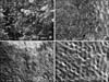
Photomicrographs for SEM observations of erupted and unerupted enamel are provided in Fig 2. Remarkable differences in the buccal enamel surfaces of erupted and unerupted teeth were observed. The surface of erupted tooth has an unclear and irregular structure. However, the unerupted tooth has a prismatic enamel structure.
DISCUSSION
Sheen et al.15 reported that bond strength inolder permanent teeth was greater than in younger teeth, regardless of etching time. Bhaskar16 found that the enamel surfaces of unerupted and recently erupted teeth are completely covered with pronounced perikymata and rod-ends. With age, the perikymata and rod-ends may wear away. As a result of time changes in the organic portion of enamel, presumably near the surface, teeth may become harder and thereby reinforce the bond strength.16 Oliver13 investigated the bond strength of orthodontic attachments to enamel from two groups of teeth (erupted premolars and unerupted canines) and concluded that the bond strength of the enamel/adhesive interface is, in fact, different for erupted and unerupted enamel. Tüfekçi et al.11 reported that there were no differences in bond strengths between teeth with mature and newly erupted enamel etched with either self-etching adhesive or conventional etching techniques. Almy2 indicated that the post-eruptive enamel maturation process may have little effect on bond strength values when etched either conventionally or with 3M Unitek self-etching primer. Using a subjective measurement of etching patterns when viewed under SEM, Nordenvall et al.17 reported that more deep retentive surfaces were obtained when conventionally etching newly erupted teeth for 15-seconds and mature teeth for 60-seconds. In the present study, due to possible effects of the post-eruptive enamel maturation process, the erupted and unerupted teeth groups were analyzed for bond strength differences and significant differences were found in two self-etching groups (Clearfil SE Bond and G Bond) between the erupted and unerupted teeth. Conventional groups' SBS values were not found statistically different and agree with previous work which also found no differences in bond strengths between erupted and unerupted teeth.13
Many orthodontic attachment base designs are in clinical use today. Sharma-Sayal et al.18 found that attachment base designssignificantly affected mean shear bond strength. Brackets with foil-mesh bases have also been shown to have higher bond strengths than those with integral milled bases.18 Additionally, a reduction in bond strength was found associated with the reduction of base surface area.19 In the present study, shear bond strength of the attachments were lower than previous studies. We thought that the lower results of this study can be explained by the attachment base design (Fig 3) and base surface area. We did not use foilmesh bases in the present study; and base surface area was lower than the conventional brackets.
Present findings of the comparisons of different adhesive systems do not agree with previous studies that have reported lower bond strength values with self-etching adhesives. No statistically significant differences were determined among 7 adhesive groups in both erupted and unerupted teeth. A self-etching adhesive, Clearfil S3 Bond showed the highest mean SBS value in unerupted teethand conventional etching/bonding systems, Prime & Bond NT and Single Bond showed the highest mean value in erupted teeth. This is somewhat in accordance with the results of Buyukyilmaz et al.20 that reported higher bond strengths with the 3M self-etching primer. The findings from the current study do agree with Dorminey et al.21 who found no difference between conventional etching and the 3M self-etching primer when used according to the manufacturer's instructions.
The ARI scores in the current study indicated that the differences in the amount of adhesive remaining on the enamel surfaces after debonding were not significant when the erupted and unerupted teeth groups were compared. The mode of bond failure of erupted and unerupted teeth was usually at the adhesive interface (at the button-resin interface-Score 3). Tüfekçi et al.11 reported that, there were significant differences in ARI scores between mature and newly erupted teeth. Newly erupted teeth had more adhesive bond failures, whereas mature teeth had more cohesive bond failures, suggesting some differences between the 2 groups in the quality of the bond formed.
The mode of bond failure in this study for both conventional and self-etching systems was usually at the adhesive interface (at the button-resin interface-Score 3) and showed no statistically significant differences. These results agree with some previous works. Bishara et al.22 found that self-etching primers left more adhesive on the teeth. Other studies claimed that less adhesive was left on the teeth in the self-etching primer group compared with the conventional group.20,23,24 These conflicting results can be attributed to the highly subjective nature of ARI scores and the fluoride content in the enamel of the teeth tested.10
According to SEM evaluations, buccal enamel surfaces of erupted and unerupted teeth revealed aspects which varied from each other. The SEM photographs confirmed that the prismatic view of the surface of an erupted tooth is lost via calcium and other mineral precipitation during post-eruptive maturation. This situation might be an advantage for strong bonding of self-etch adhesives to unerupted enamel surfaces. Self-etching adhesives do not require a separate acid-etch step. They are composed of aqueous mixtures of acidic functional monomers, generally phosphoric acid esters, with a pH relatively higher than that of phosphoric acid-etching gels.25 Thus, self-etching adhesives do not etch enamel to the level obtained with phosphoric acid26 on erupted tooth surfaces. However, because unerupted teeth lack post-eruptive maturation, self-etching systems may optimize the etching of unerupted enamel to the level obtained with phosphoric acid.
CONCLUSION
After our encouraging laboratory findings and having in mind all the shortcomings of an in vitro setting we concluded that:
The SBS values between erupted and unerupted teeth were not significantly different between each other, except for two self-etching adhesives (Clearfil SE (Bond and G Bond).
Among investigated adhesive systems, there were no differences in SBSs between teeth that were prepared for bonding with conventional and self-etching systems.
For both tooth types (erupted and unerupted) and adhesive systems (conventional and self-etching) the distribution of the ARI scores indicated that bond failures were more frequently at the button-adhesive interface.


 XML Download