

 PDF
PDF ePub
ePub Citation
 Print
Print


Abstract
Objective
The objective of this study was to evaluate the displacement pattern and the stress distribution of the finite element model 3-D visualization during symphyseal widening according to the osteotomy position, osteotomy type, and distraction device.
Methods
The kinds of distraction devices used were tooth-borne type, hybrid type, bone-borne type and tooth-borne type 30° angulated, and the kinds of osteotomy design were vertical osteotomy line between the central incisors and step osteotomy line through the symphysis.
Results
All reference points of the mandible including the condyles were displaced laterally irrespective of the osteotomy position, osteotomy method and distraction device. The anteroposterior or vertical displacements showed small differences between the groups. The widening pattern of the osteotomy line in the tooth-borne type of device was v shaped, and that of bone-borne type was a reverse v shape. However, the pattern in the hybrid type was parallel. The lateral displacement of the mandibular angle by the bone-borne device was more remarkable than the other types of devices. The displacement by the 30° angulated tooth-borne type was different between the left and right sides in both the transverse and anteroposterior aspects.
Figures and Tables
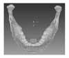
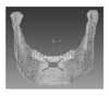
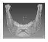
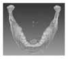
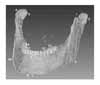
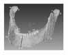
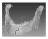
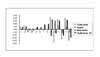
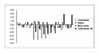
Fig 11
Displacement contour of experimental group 1 (tooth borne type in vertical osteotomy between central incisors)
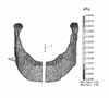
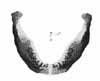
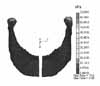
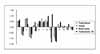

Table 2
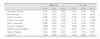
Mechanical properties of tooth, periodontal membrane, cortical bone, trabecular bone, and distraction device

Table 3
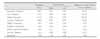
Direction of muscular orthogonal components derived as unit vectors (i.e., direction cosines) from single vectors of muscular attachment


References
1. Kim YW, Sohn BH. A study on initial changes during canine retraction by the finite element method. Korean J Orthod. 1988. 18:25–53.
2. Creekmore TD, Eklund MK. The possibility of skeletal anchorage. J Clin Orthod. 1983. 17:266–269.
3. Proffit WR, White RP. Surgical-orthodontic treatment. 1990. St Louis: Mosby.
4. Chin M, Toth BA. Distraction osteogenesis in maxillofacial surgery using internal device: Review of five cases. J Oral Maxillofac Surg. 1996. 54:45–53.

5. Kim SC, Tae KC. Corticotomy and the intrusive Tooth Movement. Korean J Orthod. 2003. 33:399–405.
6. Liou EJ, Huang CS. Rapid canine retraction through distraction of periodontal ligament. Am J Orthod Dentofacial Orthop. 1998. 114:372.
7. Tae KC, Kang KH, Min SK. Case reports of antero-posterior movement with distraction osteogenesis in maxillary anterior segment. Korean Assoc Maxillofac Plast Reconstr Surg. 2003. 25:358–363.
8. Gosain AK. Distraction osteogenesis of the craniofacial skeleton. Plast Reconstr Surg. 2001. 107:278–280.
9. Lynch SE, Genco RJ, Marx RE. Tissue engineering. Quintessence. 1999.
10. McCarthy JG, Stelnicki EJ, Mehrara BJ, Longaker MT. Distraction osteogenesis of the craniofacial skeleton. Plast Reconstr Surg. 2001. 107:1812–1827.
11. Altuna G, Walker DA, Freeman E. Surgically assisted rapid orthodontic lengthening of the maxilla in primate? A pilot study. Am J Orthod Dentofacial Orthop. 1995. 107:531–536.
12. Figueroa AA, Polley JW. Management of the sever cleft maxillary defiency with distraction osteogenesis: Procedure and result. Am J Orthod Dentofacial Orthop. 1999. 115:1–20.
13. Bell WH, Epker BN. Surgical orthodontic expansion of the maxilla. Am J Orthod. 1976. 70:517–528.
14. Smith SW, Sachdeva RC, Cope JB. Evaluation of the consolidation period during osteodistraction using computed tomography. Am J Orthod Dentofacial Orthop. 1999. 116:254–263.
15. Del Santo M Jr, English JD, Wolford LM, Gandini LG Jr. Midsymphyseal distraction osteogenesis for correcting transverse mandibular discrepancies. Am J Orthod Dentofacial Orthop. 2002. 121:629–638.
16. Proffit WR, Henry W, Fields JR. Contemporary orthodentics. 2000. St Louis: Mosby.
17. Park YC, Hwang HS, Choi KC. Biomechanics in clinical orthodontics. 1997. Seoul: Narae Publishing.
18. Contasti G, Guerrero C, Rodriguez AM, Legan HL. Mandibular widening by distraction osteogenesis. J Clin Orthod. 2001. 35:165–173.
19. Hollis BJ, Block MS, Gardiner D, Chang A. An experimental study of mandibular arch widening in the dog using distraction osteogenesis. J Oral Maxillofac Surg. 1998. 56:330–338.
20. Weil TS, Van Sickels JE, Payne CJ. Distraction osteogenesis for correction of transverse mandibular deficiency: A preliminary report. J Oral Maxillofac Surg. 1997. 55:953–960.
21. Guerrero CA, Bell WH, Rodriguez AM. Mandibular widening by intraoral distraction osteogenesis. Br J Oral Maxillofac Surg. 1997. 35:383–392.
22. Cope JB, Samchukov ML, Cherkashin AM. Mandibular distraction osteogenesis: A historic perspective and future direction. Am J Orthod Dentofacial Orthod. 1999. 115:448–460.
23. Von Sickels JE. Distraction osteogenesis versus orthognathic surgery. Am J Orthod Dentofacial Orthod. 2000. 118:482–484.
24. Mommaerts MY. Bone anchored intraoral device for transmandibular distraction. Br J Oral Maxillofac Surg. 2001. 39:8–12.
25. Del Santo M Jr, Guerrero CA, Buschang PH, English JD, Samchukov ML, Bell WH. Long-term skeletal and dental effects of mandibular symphyseal distraction osteogenesis. Am J Orthod Dentofacial Orthop. 2000. 118:485–493.
26. Conley R, Legan H. Mandibular Symphyseal Distraction Osteogenesis: Diagnosis and Treatment Planning Considerations. Angle Orthod. 2003. 73:3–11.
27. King JW, Wallace JC, Scanlan D. A new appliance for mandibular widening by distraction osteogenesis. J Clin Orthod. 2001. 35:666–672.
28. Kim SC, Min SK, Oh SH, Tae KC, Kang KH. Surgically assisted rapid tooth movement. 2004. Seoul: Myungmun Publishing.
29. Park CK, Yang WS. A three-dimensional finite element analysis on the location of center of resistance during intrusion of upper anterior teeth. Korean J Orthod. 1997. 27:259–272.
30. Choi CK. Finite element method. 2002. Taejon: Techno-press.
31. Lee JY. Finite element method. 2004. Seoul: Munundang.
32. Korioth TW, Hannam AG. Deformation of the human mandible during simulated tooth clenching. J Dent Res. 1994. 73:56–66.
33. Graber TM, Vanarsdall RW. Orthodontics: Current principles and techniques. 1994. St Louis: Mosby.
34. Kim HS, Nahm DS. A finite element and strain gauge analysis on the displacement of craniofacial complex with cervical headgear. Korean J Orthod. 1987. 17:185–197.
35. Wheeler RC. A textbook of dental anatomy and physiology. 1965. Philadelphia: WB Saunders.
36. Andrews LF. The six keys to normal occlusion. Am J Orthod. 1972. 62:296–309.
37. Dewel BF. Clinical observations on the axial inclination of teeth. Am J Orthod. 1949. 35:98–115.
38. Kim JS, Jin KH, Hong SJ. A statistical study of clinical crown inclination in Korean's naturally occuring optimal occlusion. Korean J Orthod. 1992. 22:715–733.
39. Choi BT, Yang WS. A roentgenocephalometric study on mesiodistal axial inclination of posterior teeth. Korean J Orthod. 1984. 14:151–160.
40. Noyes HJ, Rushing CH, Sims HA. The angle of axial inclination of human central incisor teeth. Angle Orthod. 1943. 13:60–61.
41. Coolidge ED. The thickness of the human periodontal membrane. J Am Dent Assoc. 1937. 24:1260–1270.
42. Tanne K, Koenig HA, Burstone CJ. Moment to force ratios and the center of rotation. Am J Orthod Dentofacial Orthod. 1988. 94:426–431.
43. Joe BJ, Sohn BH. A finite element analysis of the stress distribution and displacement in human maxilla to rapid palatal expansion. Korean J Orthod. 1985. 15:43–54.
44. McGuinness N, Wilson AN, Jones M, Middleton J, Roberson NR. Stresses induced by edgewise appliances in the periodontal ligament - a finite element study. Angle Orthod. 1992. 62:15–22.
45. Kim MK. Anatomy of head and neck. 1998. Seoul: Medical dental publishing.
46. Kim JH, Chung JH, Cho KZ. Finite element analysis of mandibular stresses and denture movements induced by overdentures. J Korean Acad Prosthodont. 1990. 28:63–94.
47. Tahk SG, Park YC. A study on craniofacial growth analysis of Korean children by the finite element method. Korean J Orthod. 1988. 18:343–366.
48. Kim JY, Sohn BH. A finite element analysis on the effect of the reverse headgear to the maxillary complex. Korean J Orthod. 1985. 15:7–22.
49. Choue HK, Lee KS. A finite element analysis of the stress distribution and displacement of an in-vitro human mandible to the orthopedic force. Korean J Orthod. 1984. 14:75–92.
50. Jang JW, Sohn BH. A study on the pattern of movement during retraction of maxillary central incisor by finite element method. Korean J Orthod. 1991. 21:617–634.
51. Chun KM, Nahm DS. Mechanical analysis of the multiloop edgewise arch wire. Korean J Orthod. 1991. 21:31–47.
52. Hwang CI, Suhr CH. Three-dimensional finite element analysis on reciprocal action by torque application in maxillary archwire. Korean J Orthod. 1994. 24:479–508.
53. Ilizarov GA. The tension-stress effect on the genesis and growth of tissues Part I. The influence of stability of fixation and soft-tissue preservation. Clin Orthop Pleat Res. 1989. 238:249–281.
54. Snyder CC, Levine GA, Swanson HM, Browne EZ. Mandibular lengthening by gradual distraction. Plast Reconstr Surg. 1973. 51:506–508.
55. Shapiro PA. Mandibular dental arch form and dimension: treatment and postretension changes. Am J Orthod. 1974. 66:58–70.
56. Gardner SD, Chaconas S. Posttreatment and postretention changes following orthodontic therapy. Angle Orthod. 1976. 46:151–161.
57. Strang RHW. The fallacy of denture expansion as a treatment procedure. Angle Orthod. 1949. 19:12–22.
58. Kisnisci RS, Fowel SD, Epker BN. Distraction osteogenesis in silver Russell syndrome to expand the mandible. Am J Orthod Dentofacial Orthod. 1999. 116:25–30.
59. Bell WH, Gonzalez M, Samchukov ML, Guerrero CA. Intraoral widening and lengthening the mandible in baboons by distraction osteogenesis. J Oral Maxillofac Surg. 1999. 57:548–562.
60. Samchukov ML, Cope JB, Cherkashin AM. The effect of sagittal orientation of the distractor on biomechanics of mandibular lengthening. J Oral Maxillofac Surg. 1999. 57:1214–1222.
61. Cope JB, Yamashita J, Healy S, Dechow PC, Harper RP. Force level and strain patterns during bilateral mandibular osteodistraction. J Oral Maxillofac Surg. 2000. 58:171–178.
62. Samchukov ML, Cope JB, Harper RP, Ross JD. Biomechanical considerations of mandibular lenthening and widening by gradual distraction using a computer medel. J Oral Maxillofac Surg. 1998. 56:51–59.
63. Harper RP, Bell WH, Hilton RJ, Browne R, Cherkashin AM, Samchukov ML. Reactive changes in the temporomandibular joint after mandibular midline osteodistraction. Br J Oral Maxillofac Surg. 1997. 35:20–25.
64. Braun S, Bottrel A, Legan HL. Condylar displacement related to mandibular symphyseal distraction. Am J Orthod Dentofacial Orthop. 2002. 121:162–166.
 XML Download