Abstract
Purpose:
The purpose of this study was to determine the correlation and ratio between the calcaneal length and width for predicting the width of calcaneus.
Materials and Methods:
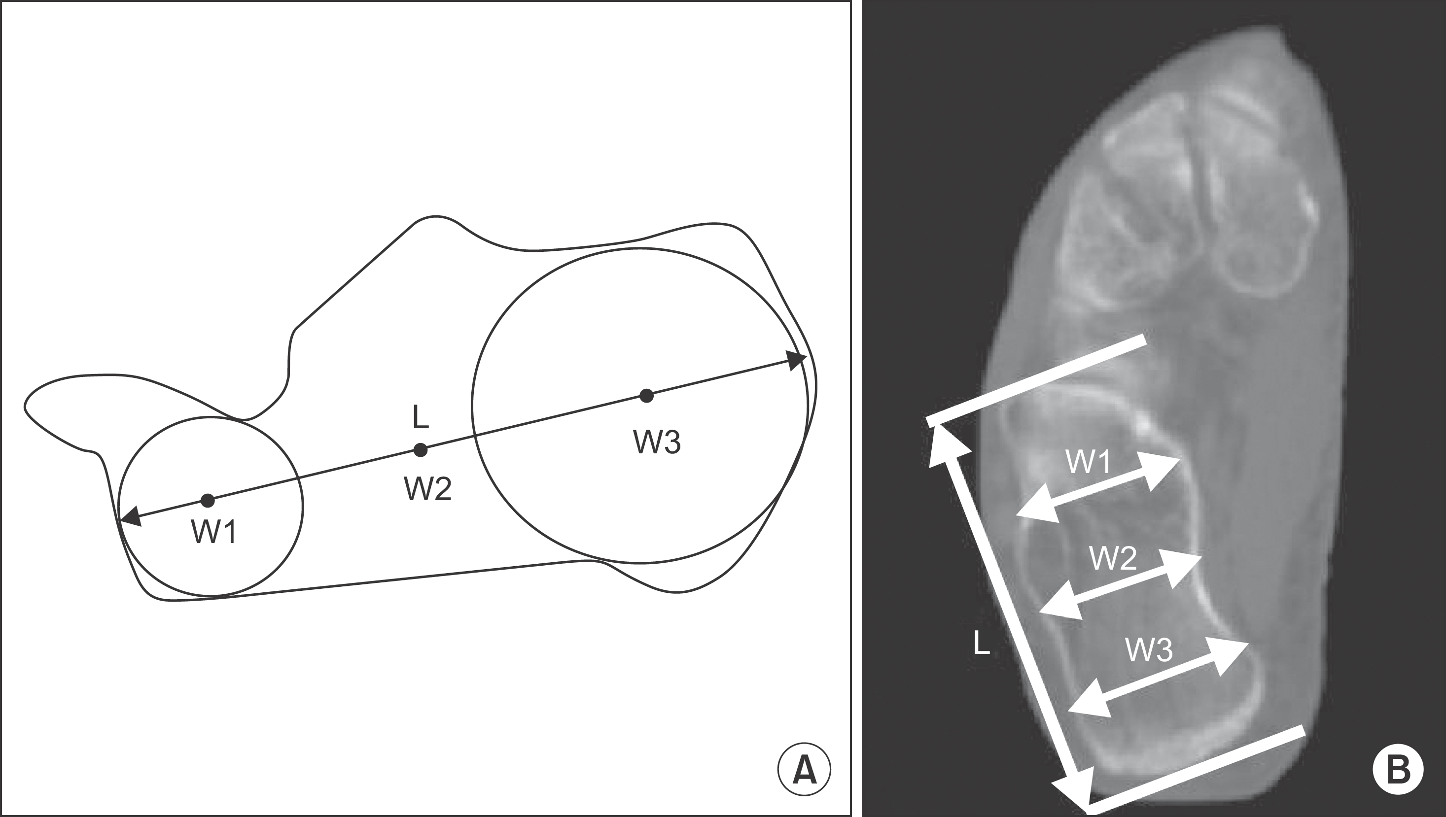
A total of 190 feet (190 patients) were included based on computed tomography scans. The length of calcaneus (CL) was measured on the line connecting the center of a circle tangent to the cortical margin in the anterior and posterior parts of the calcaneus in a sagittal plane (W1, W2). The width of the calcaneus was defined as the horizontal line of each part (W1, W2, W3) on the same axial plane. The relationship between the measurement was determined through a correlation analysis. The reliability was assessed based on intraclass correlation coefficients.
Results:
The CL and widths of calcaneus (W1, W2, W3) had a good positive correlation (r=0.848 [W1/CL], r=0.738 [W2/CL], r=0.769 [W3/CL]; p<0.001). The mean CL and widths ratios were 0.33 (W1/CL), 0.37 (W2/CL), and 0.37 (W3/CL). Using these ratios to estimate the widths by multiplying each ratio by the measured calcaneal length, we found a difference between the estimated calcaneal widths and the actual measured calcaneal widths values was 0.25 mm, 0.43 mm, and 0.16 mm. All measurements showed good-to-excellent inter- and intraobserver reliability.
REFERENCES
1.Biggi F., Di Fabio S., D’Antimo C., Isoni F., Salfi C., Trevisani S. Percutaneous calcaneoplasty in displaced intraarticular calcaneal fractures. J Orthop Traumatol. 2013. 14:307–10.

2.Labronici PJ., Reder VR., de Araujo Marins Filho GF., Pires RE., Fernandes HJ., Mercadante MT. Risk of injury to vascular-nerve bundle after calcaneal fracture: comparison among three techniques. Rev Bras Ortop. 2011. 51:208–13.

3.Kwon JY., Zurakowski D., Ellington JK. Influence of contralateral radiographs on accuracy of anatomic reduction in surgically treated calcaneus fractures. Foot Ankle Int. 2015. 31:75–82.

4.Basile A. Operative versus nonoperative treatment of displaced intra-articular calcaneal fractures in elderly patients. J Foot Ankle Surg. 2010. 49:25–32.

5.Qiang M., Chen Y., Zhang K., Li H., Dai H. Effect of sustentaculum screw placement on outcomes of intra-articular calcaneal fracture osteosynthesis: a prospective cohort study using 3D CT. Int J Surg. 2015. 19:72–7.

6.Santi MD., Botte MJ. External fixation of the calcaneus and talus: an anatomical study for safe pin insertion. J Orthop Trauma. 1991. 10:487–91.

7.Kendoff D., Citak M., Gardner M., Kfuri M Jr., Thumes B., Krettek C, et al. Three-dimensional fluoroscopy for evaluation of articular reduction and screw placement in calcaneal fractures. Foot Ankle Int. 2007. 28:1115–71.

8.Swanson SA., Clare MP., Sanders RW. Management of intra-articular fractures of the calcaneus. Foot Ankle Clin. 2008. 13:159–78. during fixation of calcaneal fractures: a technique tip. Foot Ankle Spec. 2014;7: 208-10.

9.David V., Stephens TJ., Kindl R., Ang A., Tay WH., Asaid R, et al. Calcaneotalar ratio: a new concept in the estimation of the length of the calcaneus. J Foot Ankle Surg. 2015. 54:370–2.

10.Ene R., Popescu D., Panaitescu C., Circota G., Cirstoiu M., Cirstoiu C. Low complications after minimally invasive fixation of calcaneus fracture. J Med Life. 2013. 1:80–3.
11.Rammelt S., Zwipp H. Calcaneus fractures: facts, controversies and recent developments. Injury. 2004. 35:443–11.

12.Abousayed MM., Toussaint RJ., Kwon JY. The use of dual C-arms during fixation of calcaneal fractures: a technique tip. Foot Ankle Spec. 2014. 7:208–10.
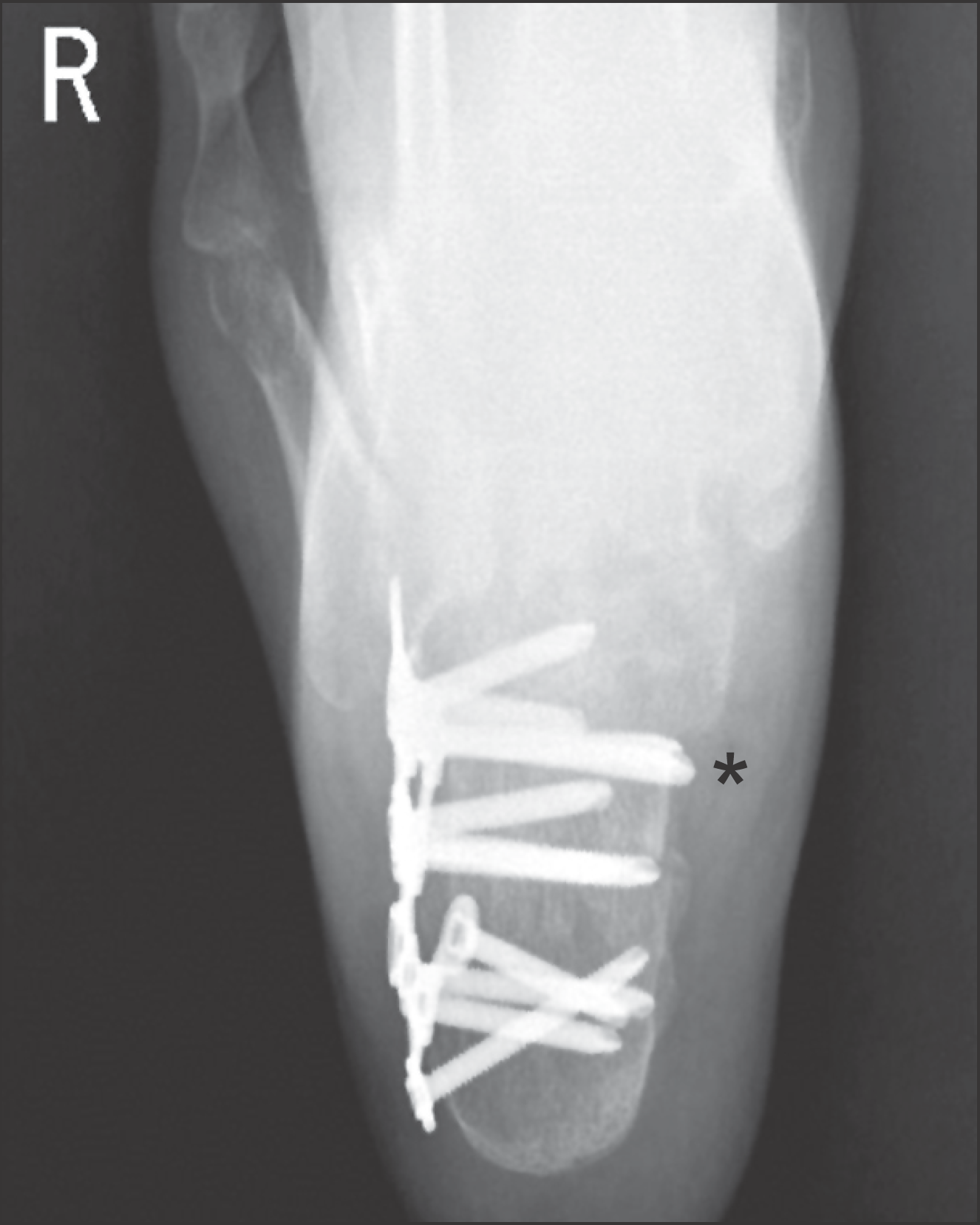
Figure 1.
Postoperative radiograph shows fixated screws of lateral to medial direction, and shows (asterisk) penetrates medial cortex of calcaneus.

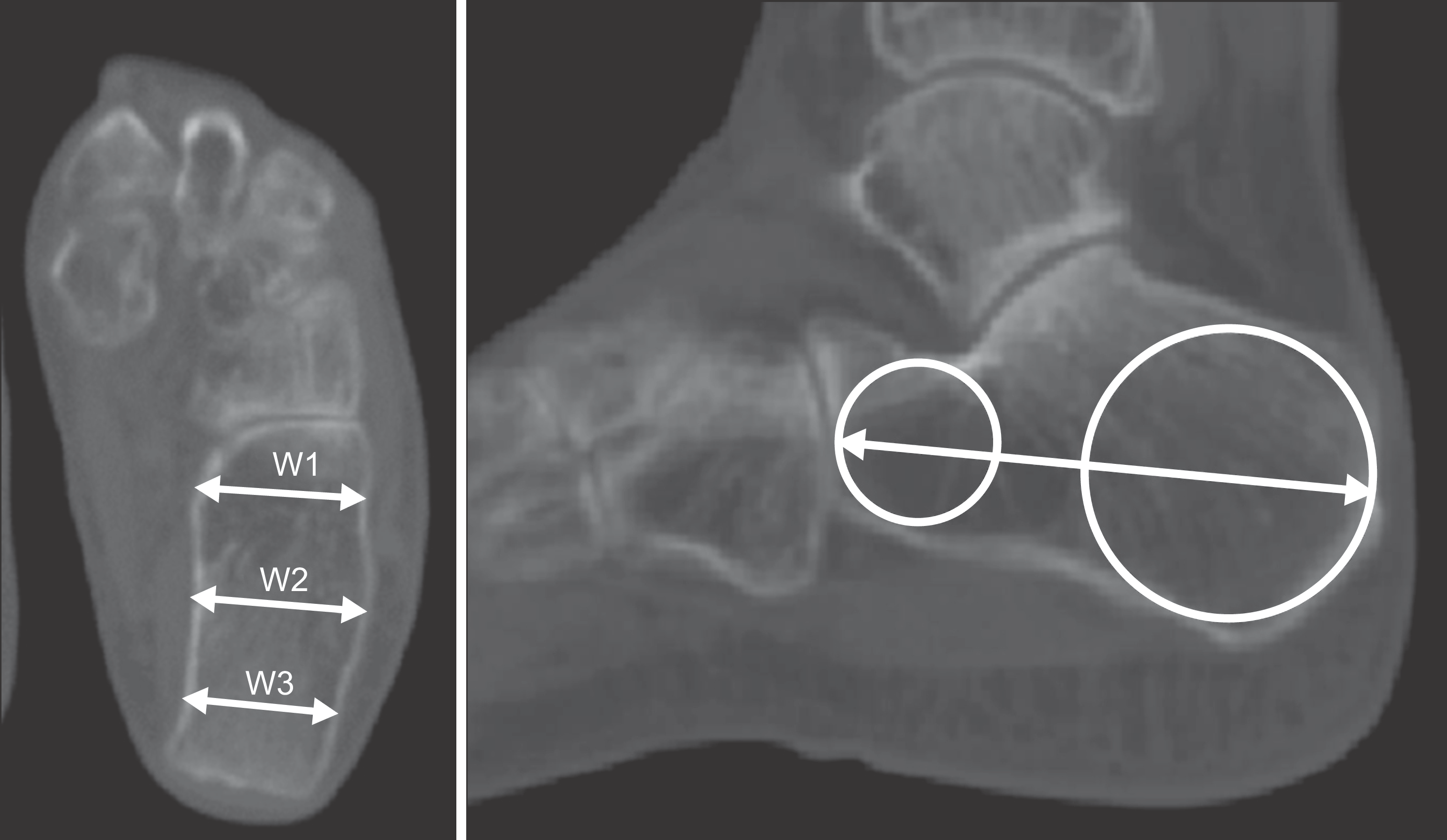
Figure 2.
Axial cutting level was decided depend on sagittal plane level involving a line connecting the center of a circle tangent to the cortical margin in the anterior (W1) and posterior (W3) parts of the calcaneus in the sagittal plane.

Figure 3.
(A) The length of calcaneus (CL) measured on the line connecting the center of a circle tangent to the cortical margin in the anterior (W1) and posterior (W3) parts of the calcaneus in the sagittal plane. (B) The width of the calcaneus is defined as horizontal line of each part (W1, W2, W3) on same axial plane. L: length.

Table 1.
Correlation between CL and Each Widths of Calcaneus
| W1 | W2 | W3 |
|---|---|---|
| CL 0.848 (p<0.001) | 0.738 (p<0.001) | 0.769 (p<0.001) |
Table 2.
Ratios of CL and Each Widths and Difference between Each Measured and Predicted Widths of Calcaneus
| W1/CL | W2/CL | W3/CL | |
|---|---|---|---|
| Ratio | 0.33±0.04 | 0.37±0.05 | 0.37±0.04 |
| Difference (mm) | 0.25±3.1 | 0.43±4.3 | 0.16±3.1 |
| IQR (Q1∼Q3) | ―1.4∼1.9 | ―1.4∼1.5 | ―2.1∼1.4 |
Table 3.
Interobserver and Intraobserver Reliability of Radiologic Measurements


 PDF
PDF ePub
ePub Citation
 Print
Print


 XML Download