

 PDF
PDF ePub
ePub Citation
 Print
Print


Abstract
Arteriovenous malformation (A-V malformation) is defined as an abnormal connection between arteries and veins that lead to A-V shunting with an intervening network of vessels. A-V malformation is a rare condition, and spontaneous regression is also rare. A-V malformation becomes symptomatic when the surrounding tissue and osseous structures are negatively affected. A-V malformation has a high recurrence rate and is relatively hard to treat. In this case, a huge mass with pulsatile and bruit on the medial plantar area were observed. With the diagnosis of A-V malformation in accordance with the results from ultrasonography, magnetic resonance imaging and computed tomography angiography, and mass excision with feeding vessel ligation through plantar midfoot approach was completed successfully.
REFERENCES
1.Shetty PC., Beninson J., Collison DW., Burke TH. Successful embolization of multiple AV foot lesions--a case history. Angiology. 1990. 41:492–7.

2.Bernard EB., Cicchinelli LD., Poindexter JM Jr. A surgical approach to a vascular malformation in the plantar medial foot. J Foot Ankle Surg. 1994. 33:37–42.
3.McNamara TO. Peripheral vascular imaging and intervention. J Vasc Surg. 1993. 18:720–1.
4.Rogalski R., Hensinger R., Loder R. Vascular abnormalities of the extremities: clinical findings and management. J Pediatr Orthop. 1993. 13:9–14.
5.Yu GV., Brarens RM., Vincent AL. Arteriovenous malformation of the foot: a case presentation. J Foot Ankle Surg. 2004. 43:252–9.
6.Kunze B., Kluba T., Ernemann U., Miller S. Arteriovenous malformation: an unusual reason for foot pain in children. Foot Ankle Online J. 2009. 2:1.
7.Kim JK., Kim JS. Arteriovenous malformation infiltrating the extensor hallucis longus tendon: a case report. J Bone Joint Surg Am1. 2010. 92:210–6.
8.Laurian C. Congenital arteriovenous malformations: what are the perspectives? Phlebolymphology. 2011. 18:149–53.
9.Hyun D., Do YS., Park KB., Kim DI., Kim YW., Park HS, et al. Ethanol embolotherapy of foot arteriovenous malformations. J Vasc Surg. 2013. 58:1619–26.
Figure 1.
(A, B) This is a photography at the first visit of outpatient deparment. There were drills and pulsation at the mass, tingling sensation on the overally entire sole aspect and sensory decease on medial plantar nerve territory (arrows). (A) Lateral view. (B) Plantar view. (C, D) Preoperative photo shows 5×4.5 cm huge mass on the medial plantar side of left foot.
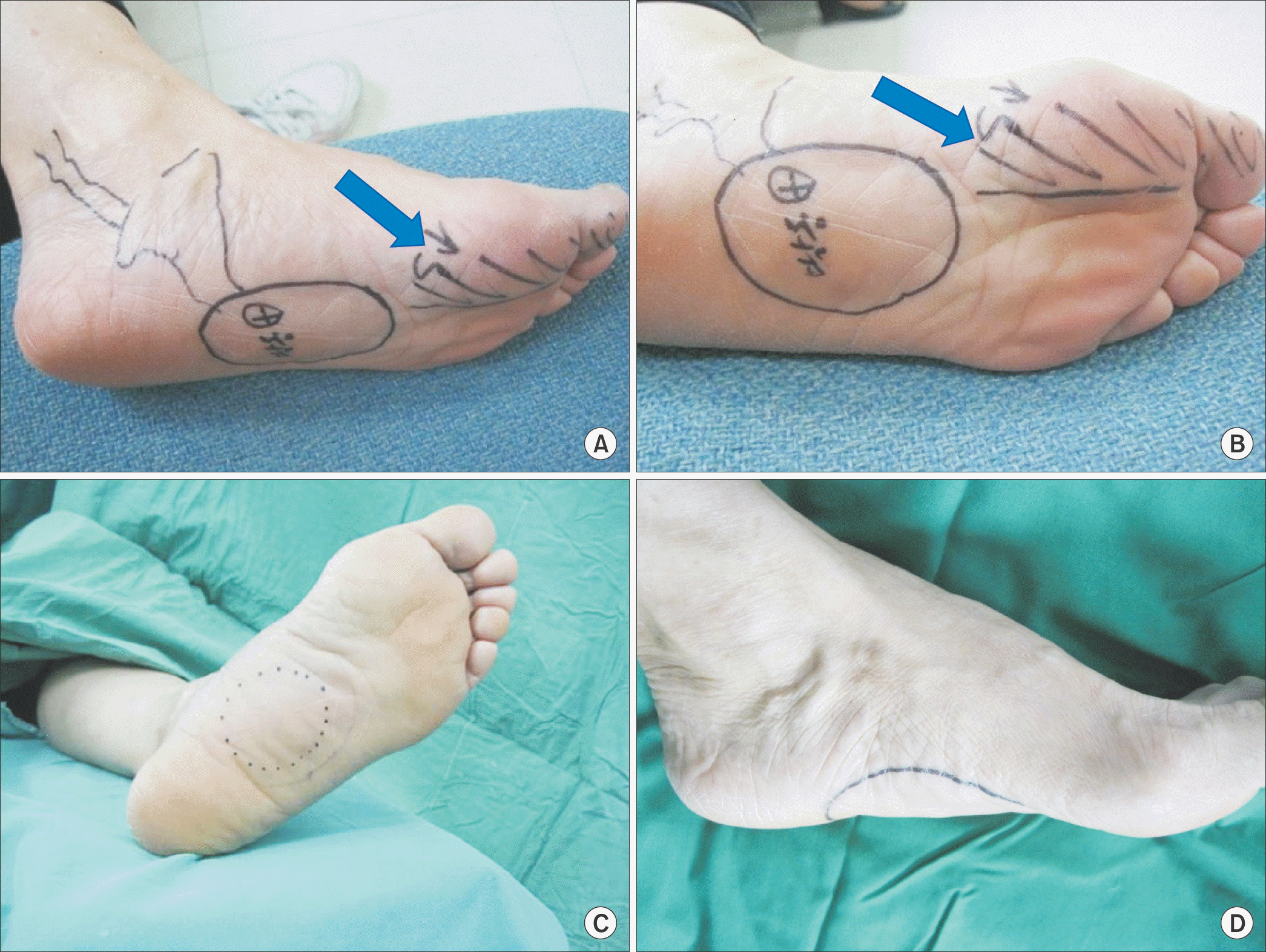
Figure 2.
Ultrasonography shows a 4.2 cm mass-like lesion of the left foot plantar surface (A), which shows extensive arterial and venous channels (B).
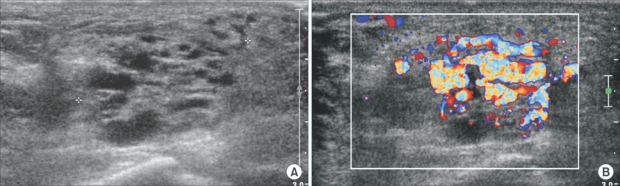
Figure 3.
Magnetic resonance imaging reveals ill-defined plantar vascular mass, mainly within flexor digitorum brevis musculature draining via enlarged posterior tibial vein. (A) Transverse view. (B) Coronal view.
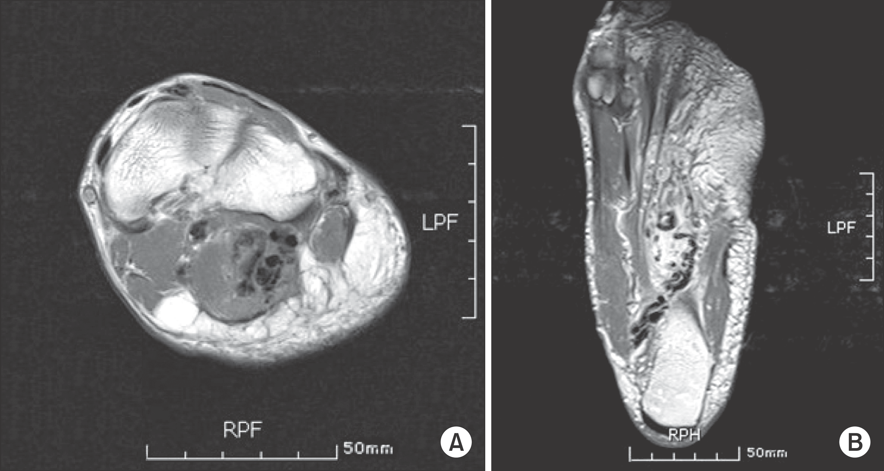
Figure 4.
(A-D) Computed tomography angiography shows that mass is mainly located in the flexor digitorum brevis (FDB) muscle, originated posterior tibial artery of midfoot around the fascia between abductor hallucis and FDB, drainged to posterior tibial vein. (E) Three-dimensional recontruction view.
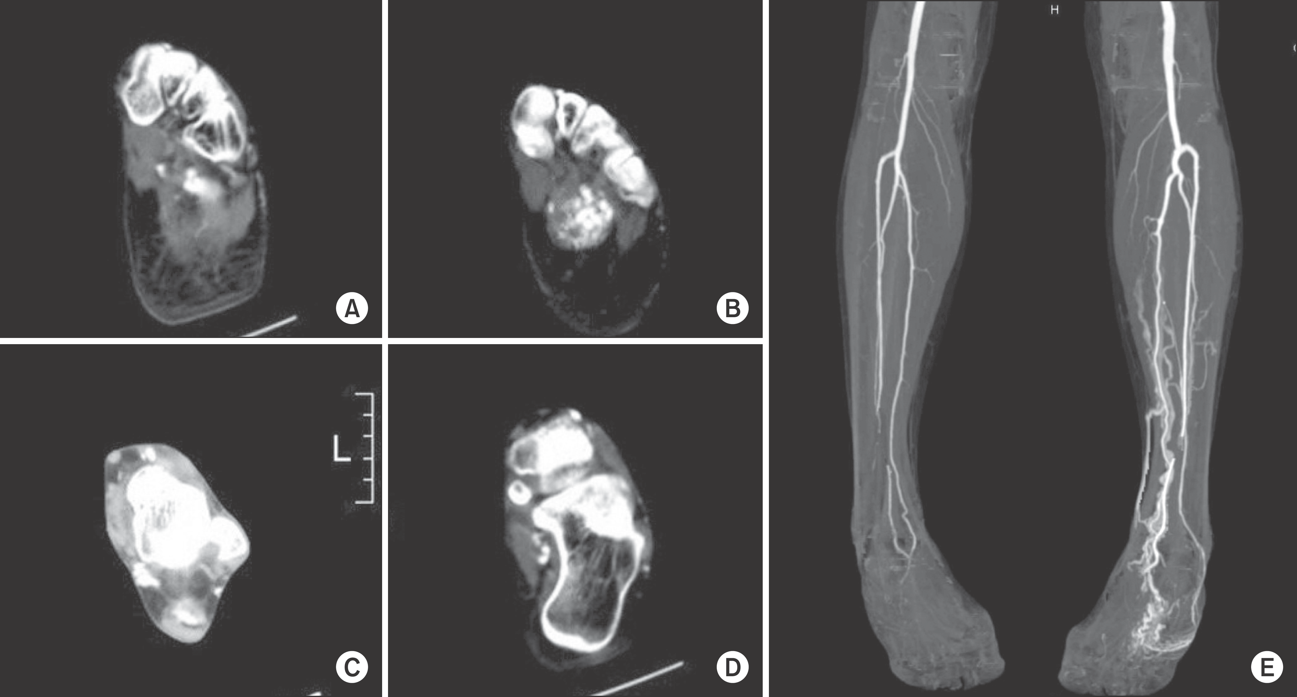
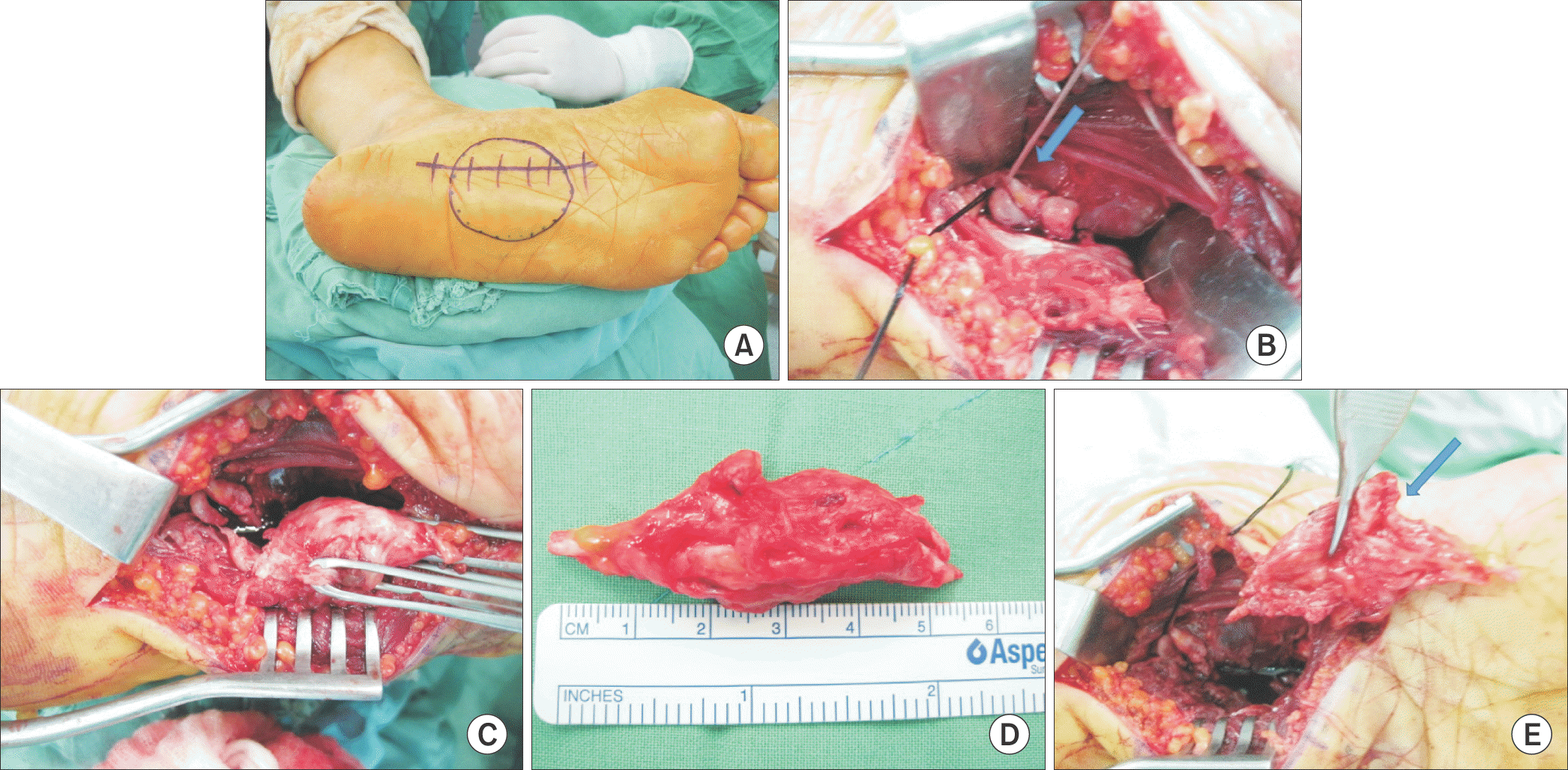
Figure 5.
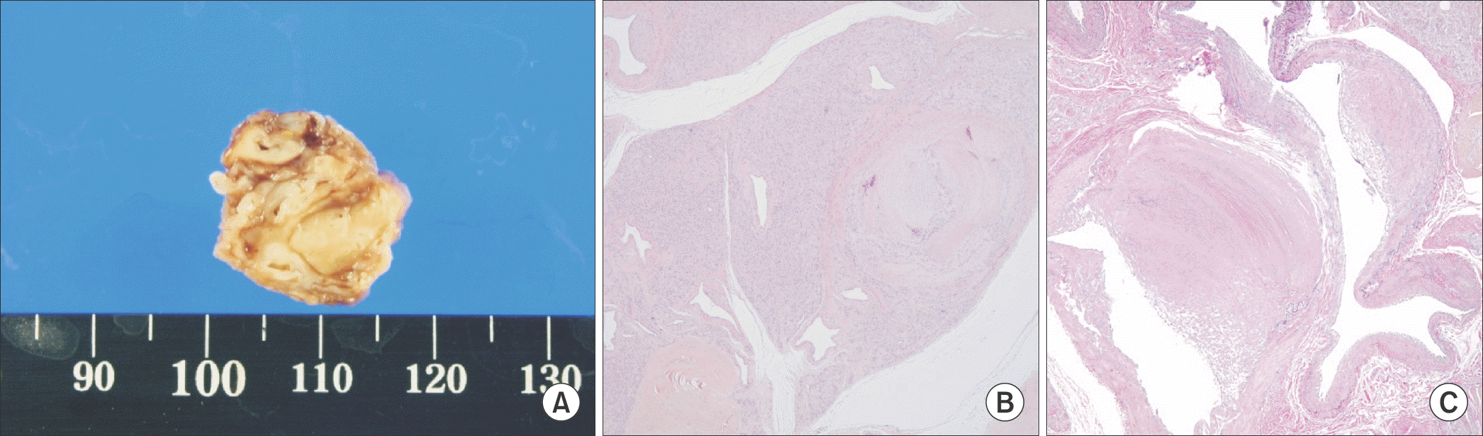
Intraoperative photography shows the procedures of the operation. Longitudinal 7 cm incision between medial aspect of plantar fascia and inferior border of abductor hallucis (A), approached to flexor digitorum brevis (FDB), incise the FDB and ligate the posterior artery and vein (arrow) (B). (C) Mass is excised. (D) Mass is measured about 5.5 cm. (E) There is fibrotic change around medial plantar nerve (arrow) due to the mass, neurolysis is applied.
 XML Download