Abstract
Spinal dural arteriovenous fistulas are rare abnormal connections of arteries and veins on the surface of the dura. A male presenting with myelopathy, which had a slowly progressive course for about 28 months, was diagnosed by magnetic resonance imaging and selective angiography. After surgical coagulation and excision, his symptoms were mildly improved. We report here on a man who underwent a surgical procedure for his myelopathy that was due to spinal arteriovenous fistula. Although it is unusual, spinal arteriovenous fistula should be considered when making a differential diagnosis of myelopathy.
REFERENCES
01). Afshar JK., Doppman JL., Oldfield EH. Surgical inter-ruption of intradural draining vein as curative treatment of spinal dural arteriovenous fistulas. J Neurosurg. 1995. 82:196–200.

02). Hurst R., Kenyon L., Lavi E. Spinal dural arteriovenous fistula: pathophysiology of venous hypertensive myelopathy. Neurology. 1995. 45:1309–1313.
03). Watson JC., Oldfield EH. The surgical management of spinal dural vascular malformations. Neurosurgery clinics of North America. 1995. 10:73–87.

04). Morimoto T., Yoshida S., Basugi N. Dural arteriovenous malformation in the cervical spine presenting with sub-arachnoid hemorrhage: a case report. Neurosurgery. 1992. 31:118–121.
05). Spetzler RF., Detwiler PW., Riina HA., Porter RW. Modified classification of spinal cord vascular lesions. J neurosurg (spine 2). 2002. 96:145–156.

06). Oldfield EH., Bennett A., Chen MY., Doppman JL. Suc-cessful management of spinal dura arteriovenous fistulas undetected by arteriography: Report of three cases. J Neurosurg. 2002. 96:220–229.
07). Rosenblum B., Oldfield EH., Doppman JL., Di Chiro G. Spinal arteriovenous malformation: a comparison of dural arteriovenous fistulas and intradural AVM's in 81 patients. J Neurosurg. 1987. 67:795–802.
08). Larsen DW., Halbach VV., Teitebaum GP., McDougall CG. Spinal dural arteriovenous fistulas supplied by branches of the internal iliac arteries. Surg Neurol. 1995. 43:35–41.

09). Larsson EM., Desai P., Hardin CW., Story J. Venous infarction of the spinal cord resulting from dural arteriovenous fistula: MR imaging findings. Am J Neuroradiol-ogy. 1991. 12:739–743.
10). Aminoff MJ., Logue V. The prognosis of patients with spinal vascular malformation. Brain. 1974. 97:211–218.
Fig. 1A-D.
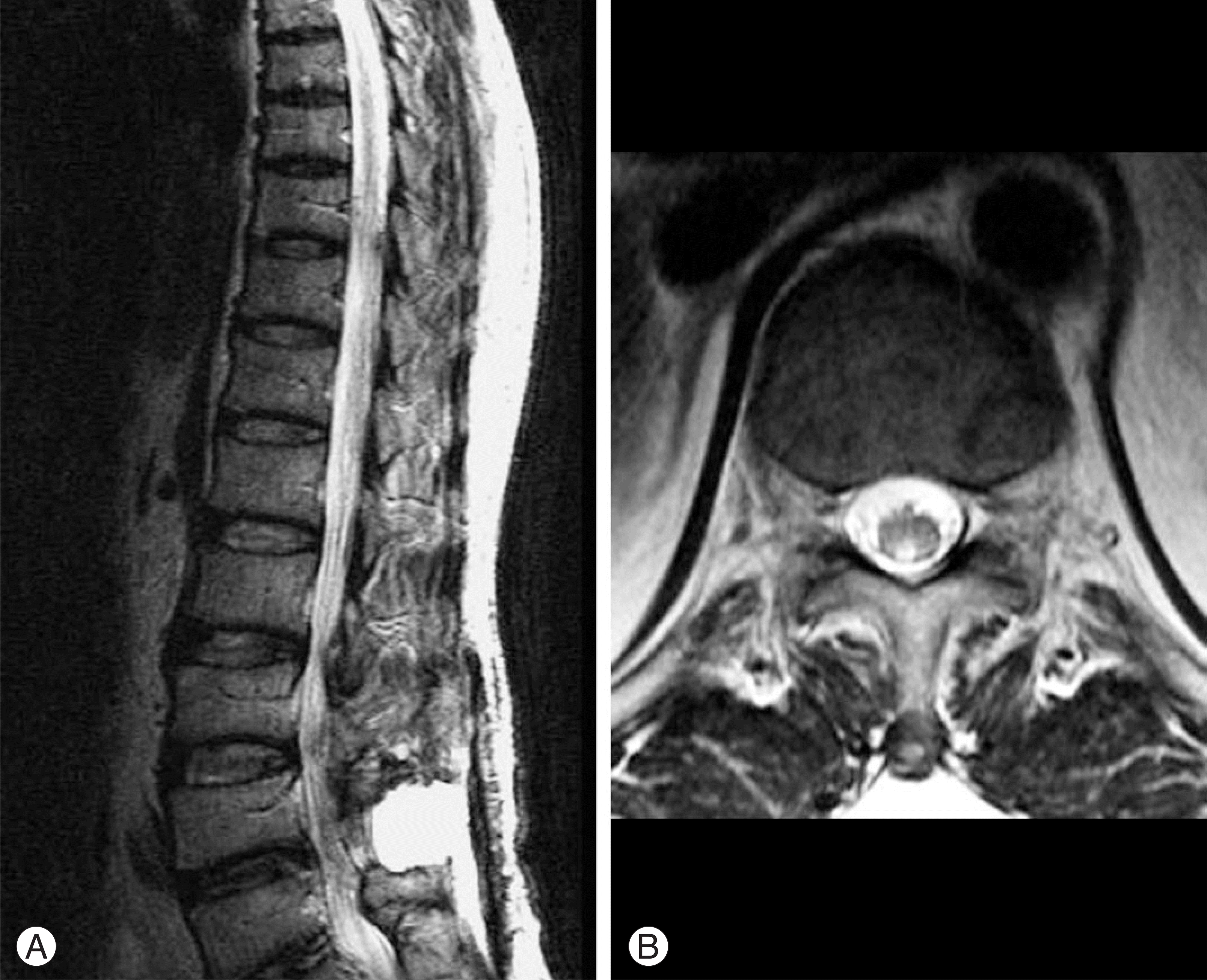
(A) Coronal and sagittal T2-weighted MRI of the thoracic spine shows marked tortuosity of the perimedullary vessel in upper and midthoracic spine level. (arrow) (B) (Right) Sagittal T2-weighted MRI of the thoracic spine shows fine dotted signal intensities in subdural space and intramedullary hyperintensity within spinal cord. (Left) Sagittal T2-weighted MRI of the thoracolumbar spine shows serpentine signal intensities within spinal cord in lumbar spine area.
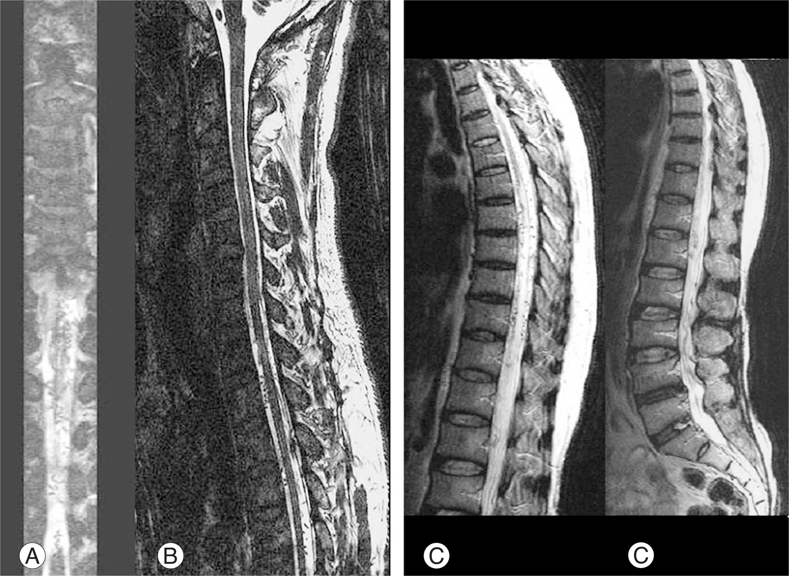
Fig. 2.
(A, B) Axial T2-weighted MRI (Right) In midthoracic level, fine dotted signal intensities in subdural space. (Left) In lumbar 3-4th disc level, abnormal dilated intradural medullary vein. (arrow)
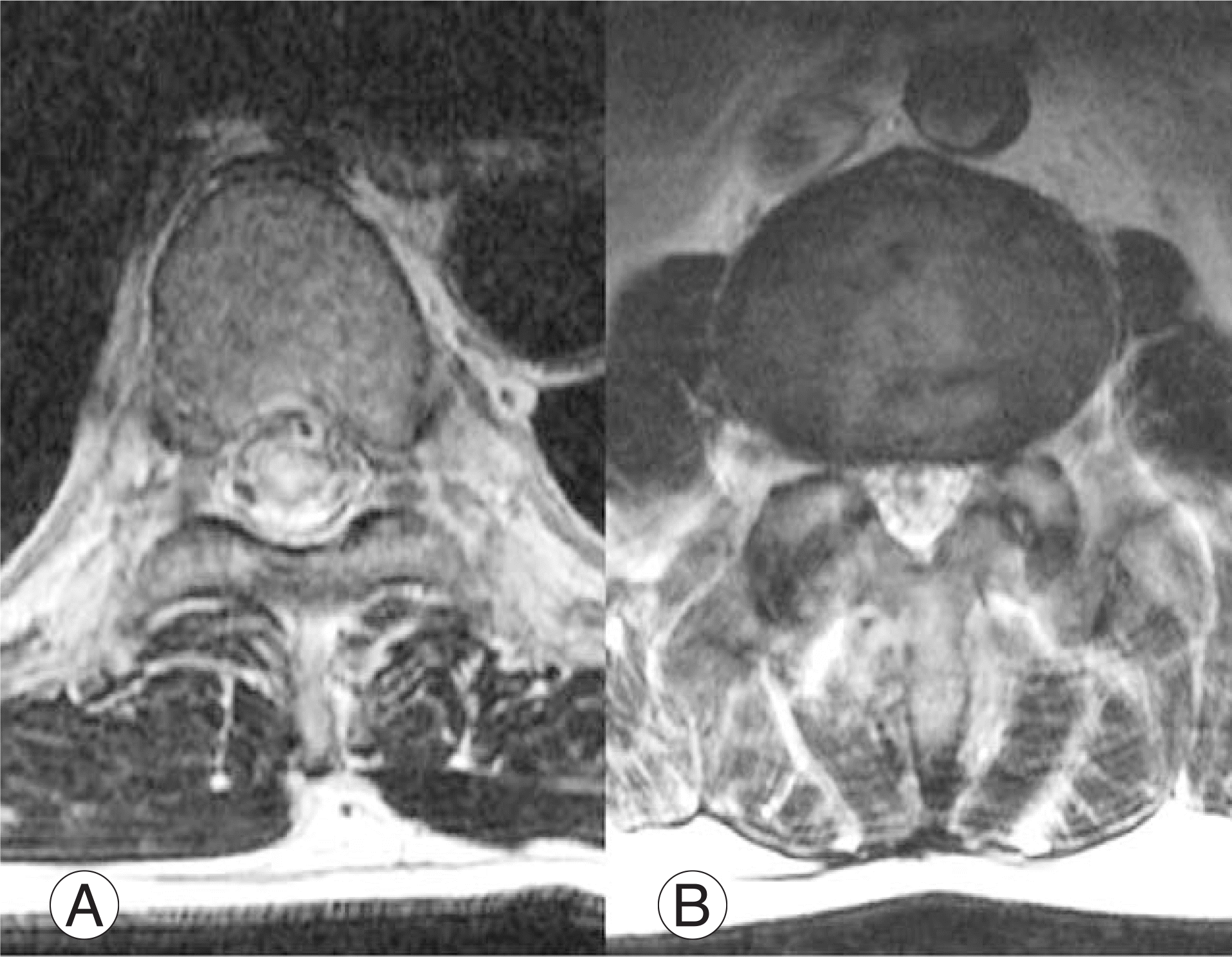
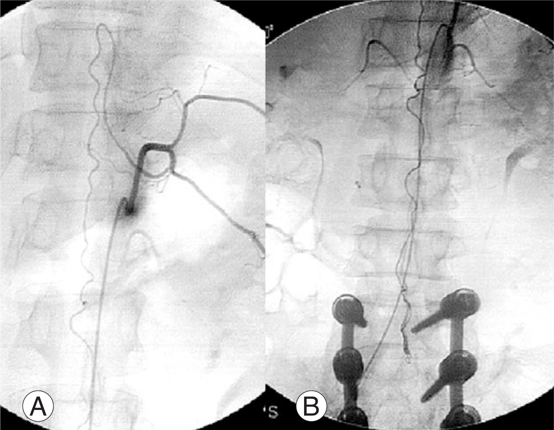
Fig. 3.
(A, B) Spinal angiography (A) feeding artery -Adamkiewicz artery comes from left thoracic 11 level. (arrow) (B) Shunt were found at lumbar 3-4th disc level. (arrow)


 PDF
PDF ePub
ePub Citation
 Print
Print


 XML Download