

 PDF
PDF ePub
ePub Citation
 Print
Print


Abstract
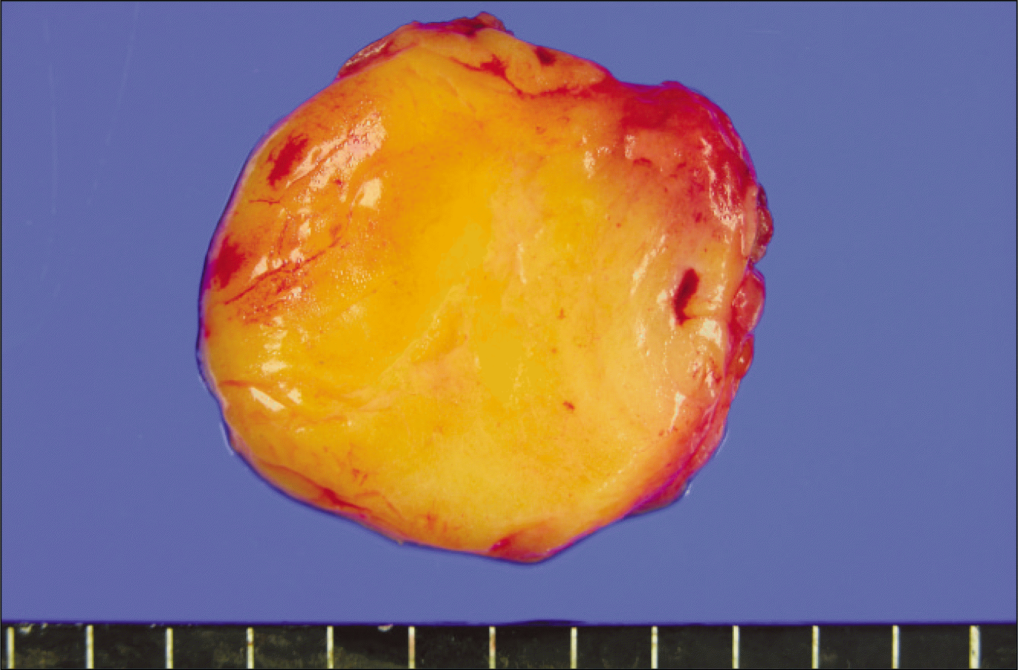
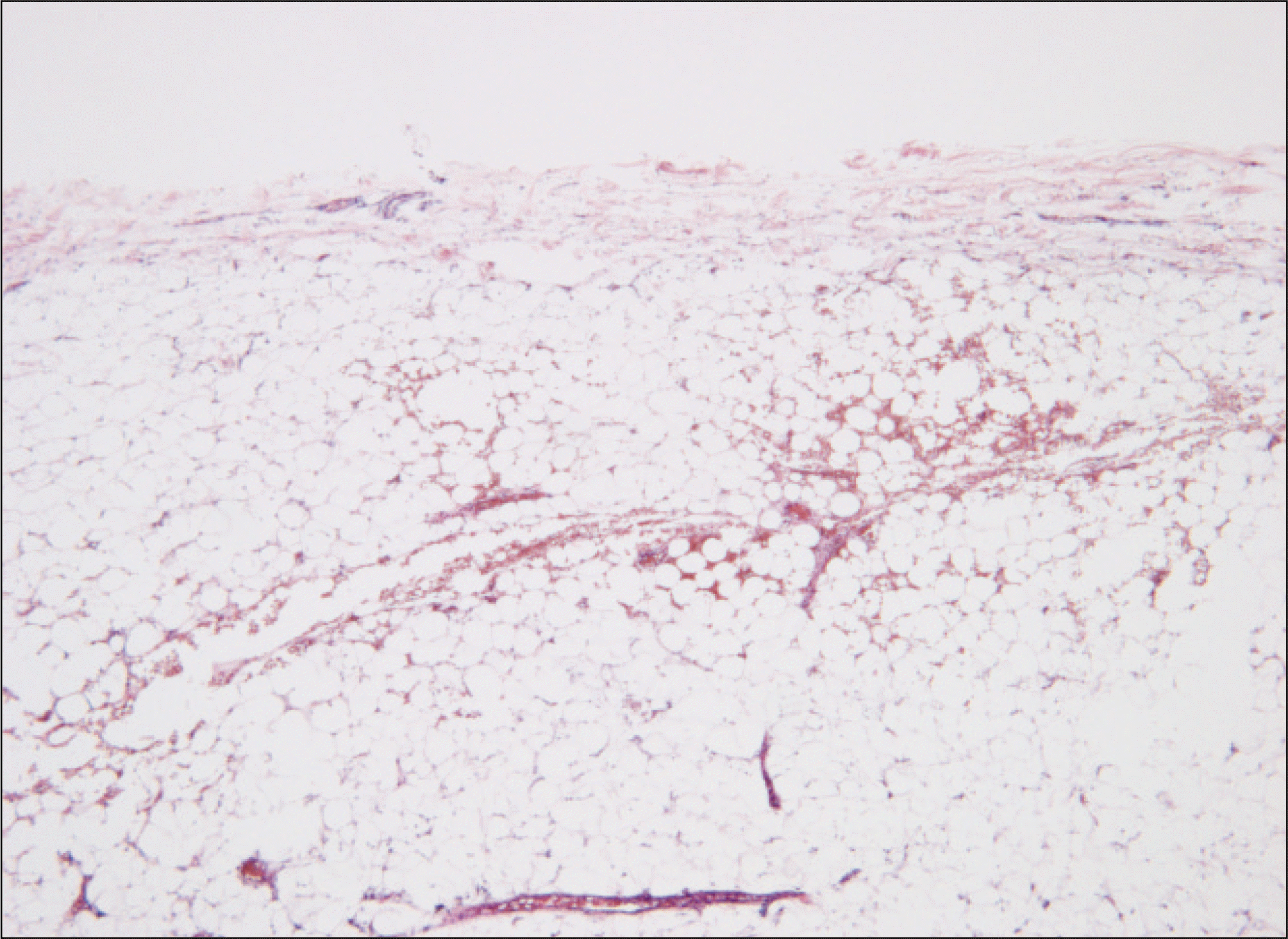
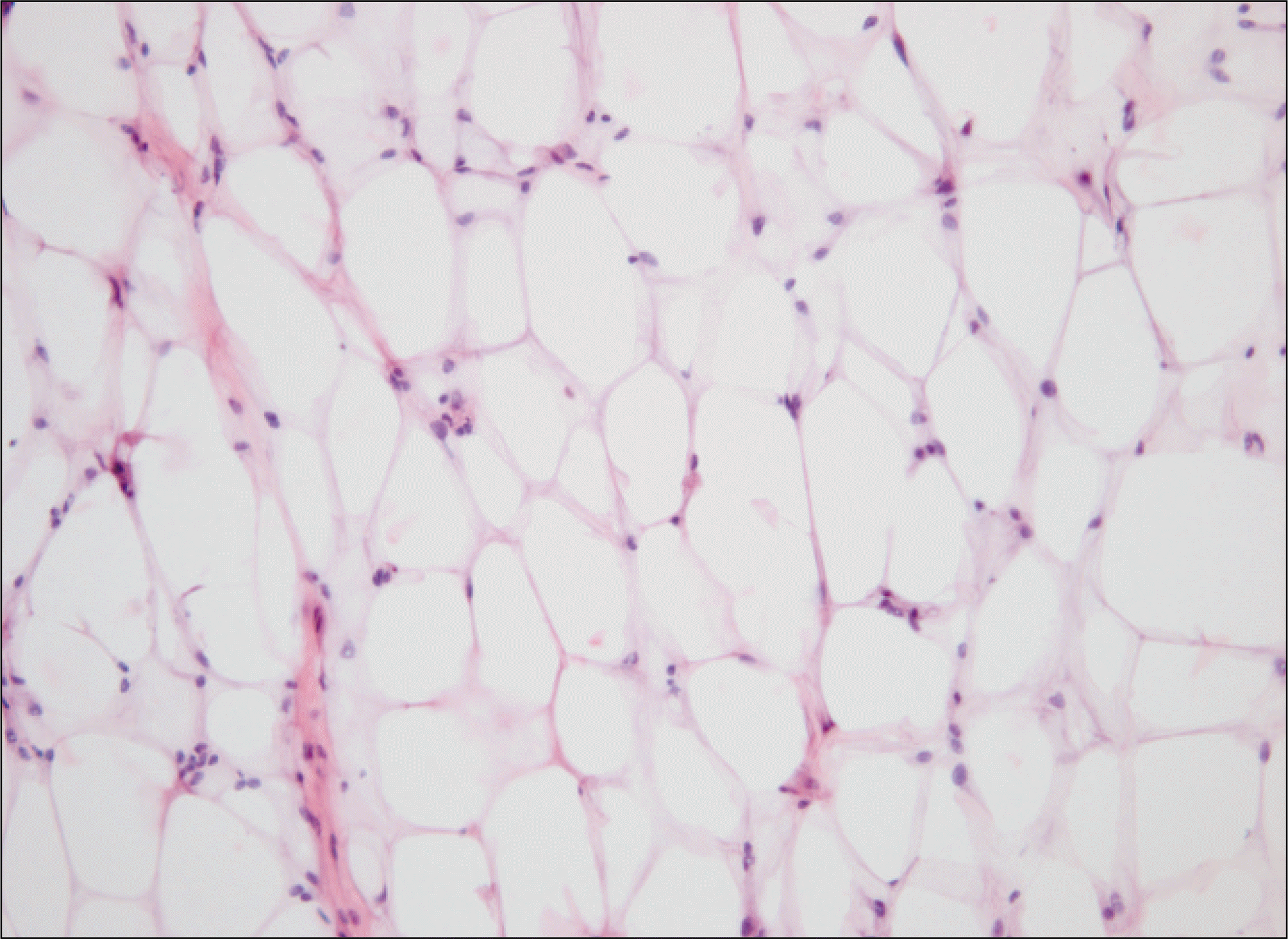
A lipoma is a benign tumor of matured adipose tissue that usually occurs at the shoulder, back, and abdomen. 13% of lipomas occur in the head and neck area. However, the incidence of lipoma in the parotid gland is very low, approximately 2.5%. A conservational surgical excision is recommended in cases of lipoma of the parotid gland, with only 1–2% of lipomas recurring. We report a case of a lipoma in the parotid gland that was removed by conservational surgical excision. The lesion was exposed by the pre-auricular approach and the tissue was detached. After the parotid gland envelop was exposed, a yellowish mass is observed that was easy to remove due to capsulation. Most authors recommend a surgical excision of the superficial lobe of the parotid gland as the treatment for a lipoma in the parotid gland. However, enucleation only may be a sufficient treatment when a lipoma occurs in the superficial lobe or around the parotid gland. A patidectomy is not needed when a lipoma is located at the superficial lobe of the parotid gland, and a conservational surgical excision is suitable. Therefore, a clinical diagnosis is important for reducing the damage to the facial nerve.
References
1. Williams TP, Stewart JC. Surgical pathology. Fonseca RJ, editor. Oral and Maxillofacial surgery. 1st ed.Philadelphia: W.B. Saunders;2000. p. 137–8.
2. Som PM, Scherl MP, Rao VM, Biller HF. Rare presentations of ordinary lipomas of the head and neck: a review. AJNR Am J Neuroradiol. 1986; 7:657–64.
3. Malave DA, Ziccardi VB, Greco R, Patterson GT. Lipoma of the parotid gland: report of a case. J Oral Maxillofac Surg. 1994; 52:408–11.

4. Walts AE, Perzik SL. Lipomatous lesions of the parotid area. Arch Otolaryngol. 1976; 102:230–2.
5. Kim YH, Reiner L. Ultrastructure of lipoma. Cancer. 1982; 50:102–6.
6. Houston GD, Brannon RB. Lipoma of the parotid gland. Oral Surg Oral Med Oral Pathol. 1985; 60:72–4.
7. Grage TB, Lober PH, Shahon DB. Benign tumors of the major salivary glands. Surgery. 1961; 50:625–33.
8. Korentager R, Noyek AM, Chapnik JS, Steinhardt M, Luk SC, Cooter N. Lipoma and liposarcoma of the parotid gland: high resolution preoperative imaging diagnosis. Laryngoscope. 1988; 98:967–71.
9. Sapp JP, Eversole LR, Wysocki GP. Contemporary oral and maxillofacial pathology. 2nd ed.St. Louis, Mo: Mosby;2004.
10. Peel RL, Gnepp DR. Diseases of the salivary glands. Barnes L, editor. Surgical pathology of the head and neck. Vol. 1. 1st ed.New York, NY: Marcel Dekker;1985. p. 533–645.
11. Janecka IP, Conley J, Perzin KH, Pitman G. Lipoma presenting as parotid tumors. Laryngoscope. 1977; 87:1007–10.
12. Enzinger FM, Weiss SW. Soft tissue tumors. 2nd ed.St. Louis, Mo: Mosby;1988.
Fig. 1.
Computed tomograph (CT) scan.(enhanced view) A. Transverse view of CT scan, B. Coronal view of CT scan.

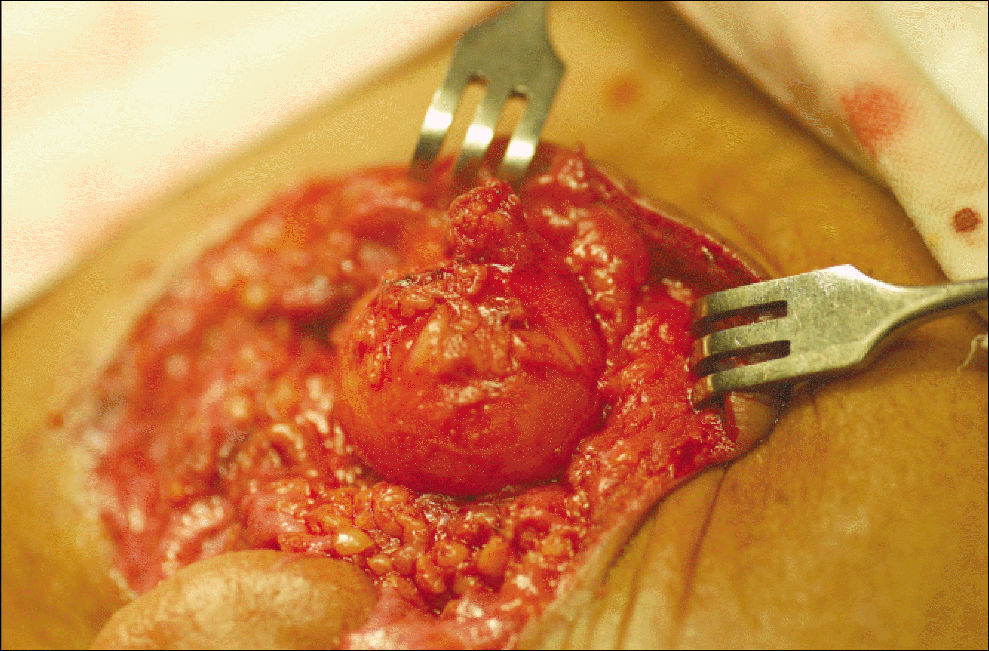
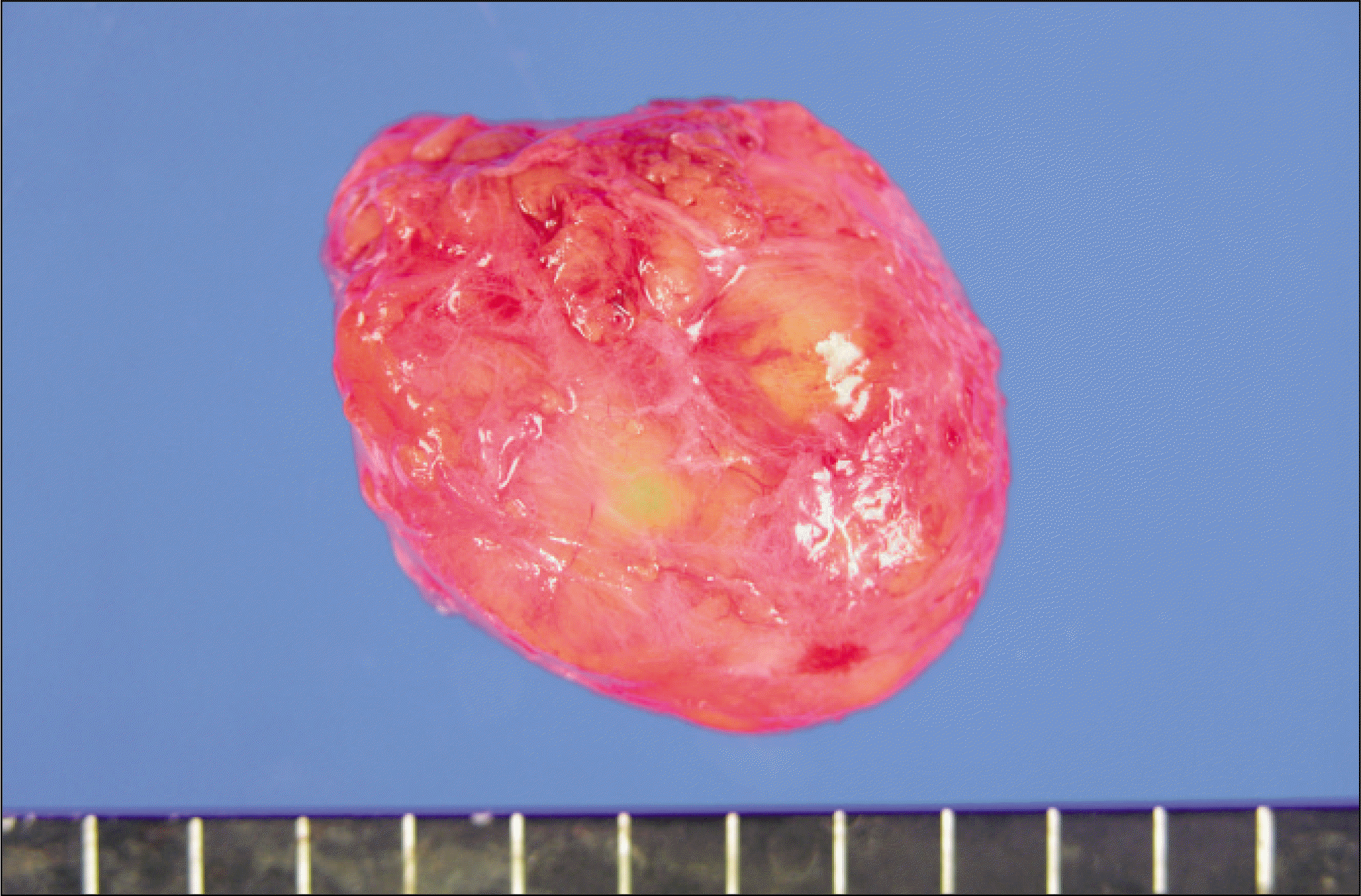
Fig. 3.
Macroscopic view (external) shows well-circumscribed, thinly encapsulated, and oval mass with soft consistency.
 XML Download