

 PDF
PDF ePub
ePub Citation
 Print
Print


Schwannomas are encapsulated tumors consisting of a pure proliferation of Schwann cells. The cells in these tumors are characteristically arranged as either solid sheets of cells (Antoni A pattern) or as stellate-to-ovoid cells in a mucinous background (Antoni B pattern). Because these tumors are cytologically benign, they require treatment only to prevent visual loss resulting from their progressive enlargement. However, it is difficult to distinguish schwannomas from amelanotic malignant melanomas. We report a case in which we diagnosed a choroidal schwannoma by surgical excision.
Case report
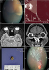
A 30-year-old woman was referred to our clinic for evaluation of an intraocular mass lesion in her right eye. Her best corrected visual acuity was 30/50 in the right eye and 20/20 in the left eye. Intraocular pressures were within normal limits for both eyes at 17 mmHg. Her ophthalmoscopic history was unremarkable except for mild myopia. Although the anterior segment showed no pathologic findings, including the absence of an afferent pupillary defect, ophthalmoscopy revealed a 3×1×1 cm-sized nonpigmented choroidal mass with subretinal fluid in the superonasal quadrant of the fundus, extending from the equator to the ora serrata (Fig. 1). The other eye was unremarkable. The mass showed relatively medium internal reflectivity and contained a cystic portion on ultrasonography (Fig. 1). Magnetic resonance imaging (MRI) showed high signal intensity in T1-weighted images with enhancement and low signal intensity in T2-weighted images (Fig. 1). A PET (18-FDG) whole body scan was performed to rule out a metastatic lesion; the scan showed no remarkable FDG uptake. We subsequently decided to perform a fine needle aspiration biopsy of the lesion with a 25G pars plana vitrectomy. The cytology showed no malignant cells. Approximately 20% of cytological biopsy results can be errorneous due to inadequate selection of the aspiration site; therefore, we did not exclusively rule out choroidal melanoma. However, because of several atypical characteristics, including the presence of cystic portions and amelanotic nonpigmented lesions, and the relatively infrequent occurrence of malignant melanoma in Korea, our differential diagnosis contained benign masses such as schwannomas and leiomyomas. Although the patient's central vision was only mildly affected, the extent of subretinal fluid could have eventually involved the macula. We decided to surgically excise the lesion to obtain a definitive diagnosis and to prevent further visual deterioration. After lamellar scleral dissection, a complete choroidal mass excision was performed by sclerouvectomy. The gross specimen was approximately 3×1×1 cm in size and contained a cystic portion (Fig. 1). Interrupted sutures were places at the scleral flaps and cryotherapy was performed at the margins of excision. A small retinal defect was found in the superonasal quadrant. Trans pars plana vitrectomy and barrier endolaser photocoagulation were performed around the retinal defect. A silicone oil injection was also performed (Fig. 1). The patient's corrected vision was 20/100 at 4 months postoperatively.
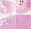
Light microscopy revealed that the tumor was composed of spindle cells with bland palisading nuclei in an Antoni A pattern. The findings were consistent with a schwannoma (Fig. 2).
Discussion
Intraocular schwannomas are very rare, benign, peripheral nerve neoplasms that usually appear as solitary, amelanotic lesions of the ciliary body or choroid. Although schwannomas are cytologically benign, they may progressively enlarge at a rate similar to or greater than that of choroidal melanomas.1 Ophthalmoscopic, MRI, and ultrasonographic findings are not helpful in differentiating schwannomas from uveal melanomas.2 In two reported cases of anterior uveal schwannomas, the tumors were noted to transilluminate brightly.3,4 This may be a helpful clinical finding because it is unusual for anterior uveal melanomas to transilluminate in this way.
Most previously reported schwannoma cases have been diagnosed after enucleations performed because of the possibility of malignant melanoma.5,6 In some reported cases, early diagnosis was made using anterior chamber biopsy techniques for ciliary body masses.7 Other cases have utilized local block excision of ciliary body tumors to confirm the diagnosis of a schwannoma.8 However, it is not known if this can always be done to confirm the presence of a schwannoma.
As was illustrated in our case, it is difficult to differentiate between an amelanotic melanoma, metastatic carcinoma, a choroidal schwannoma, or a medulloepithelioma using ancillary techniques. Immunohistochemical staining techniques were not utilized in this case. When clinical features exist that are not typical of melanoma, such as the presence of cystic components or amelanotic nonpigmented lesions (amelanotic melanoma is very rare in Korea), we recommend performing an aspiration biopsy or even surgical excision to first rule out the presence of a benign neoplasm before performing enucleation.
 XML Download