

 PDF
PDF ePub
ePub Citation
 Print
Print


Enucleation is a surgical procedure performed to treat ophthalmologic diseases, such as intraocular tumor, to relieve eyeball pain in blind eyes, or to improve disfiguring blind eye cosmesis. Enucleation consists of eyeball removal and maintenance of eyeball volume by orbital implant insertion. Porous orbital implants, such as porous hydroxyapatite and porous polyethylene (Medpor®), are most commonly elected because they allow fibrovascular ingrowth into the porous orbital implant, thus minimizing implant extrusion. Further, these implants offer high biocompatibilities, enabling extraocular muscles to be sutured directly onto implants, thus providing excellent motility.1,2
The major complication associated with porous polyethylene orbital implant use is implant exposure. For example, Karcioglu et al.3 reported Medpor® exposure in 8 of 37 retinoblastoma patients (21.6%), and Lee et al.4 reported exposure in 8 of 13 eyes (53%).
These earlier authors suggested that friction between a poorly fitting prosthesis and the tissue covering the anterior surface is the probable cause of Medpor® exposure in postenucleation retinoblastoma patients. We postulated that adding tissue between an implant and prosthesis might prevent porous polyethylene orbital implant exposure.5 The authors chose orbital fat as a buffering tissue, placing it between the Medpor® and the overlying conjunctivae, In this study, 39 orbits of retinoblastoma patients who received enucleation and Medpor® implantation in combination with free orbital fat grafts, showed no Medpor® exposure.6
The literature reports discrepancies, regarding the sustainability and survival of freely grafted orbital fat. The most accurate means of determining the viability of free orbital fat would be to graft orbital fat into human anophthalmic orbits and to follow the graft by biopsy and histological examination. However, such experiments in humans are not ethical, and biopsy of grafted fat in patients is not feasible. Therefore, the authors conducted this experiment in an animal model in order to investigate the long term survival of freely grafted orbital fat on Medpor® implants.
Materials and Methods
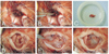
Eight adult New Zealand white rabbits were anesthetized with intramuscular ketamine hydrochloride (40 mg/kg) and xylazine (6 mg/kg). The right eye of each rabbit was initially sterilized with betadine and the eyelids were retracted with a lid speculum. A 360-degree peritomy was performed close to the corneal limbus, and the four quadrants between the rectus muscles were dissected bluntly to separate Tenon's capsule from the globe. All four rectus muscles were isolated individually with a muscle hook, secured with 6-0 polygalactin sutures, and disinserted from the globe. The optic nerve was then transected and the globe removed. Retrobulbar orbital fat (ca. 4×4×1 mm) was excised from the central portion of the posterior orbit, behind the surgically opened posterior Tenon's capsule, and stored in saline. In order to ensure that the amount of excised fat tissue was uniform, sections were weighed, using an analytical balance (ca. between 23 and 31 mg/section). After obtaining hemostasis using epinephrine-soaked gauze, Medpor® (12 mm) was placed within the intraconal space. The four recti were then sutured directly onto the anterior surface of the Medpor® at a distance of 8 mm apart. The excised orbital fat was then placed onto the anterior surface of the Medpor® an' Tenon's capsule was closed horizontally over the grafted fat, using a continuous 6-0 polygalactin suture. The conjunctiva was closed meticulously with a continuous 6-0 polygalactin suture. A conformer was then placed and tarsorrhaphy performed (Fig. 1).
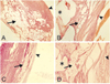
Two rabbits were randomly sacrificed at 2, 4, 8 and 12weeks post-surgery by intravenous KCL subsequent to intramuscular anesthesia. From eacy animal, the right orbits including conjunctivae, Tenon's layers and orbital implants, were excised, and all specimens were fixed in 10% formalin, decalcified, and bisected in the anteroposterior direction with a cutting plane in grafted orbital fat so that the fat can be seen in the bisected surface. The bisected implants were imbedded in paraffin for histologic examination. Sections (3 µm) were cut, stained with hematoxylin and eosin, and examined under a light microscope.
Results
Grafted orbital fat was well-maintained in the space between conjunctivae and Medpor® at 2 and 4 weeks post-surgery, but, at 8 weeks, the volume of grafted fat were significantly reduced. In addition, chronic cellular type inflammatory response was observed near the grafts. At 12 weeks post-surgery, the grafted orbital fat was composed predominantly of microcysts, with an observed loss of normal architecture and adipocytes. At this time point, no viable fat was found by histologic examinations (Fig. 2).
Discussion
Orbital fat, located behind the posterior Tenon's layer, is difficult to surgically approach and harvest. However, after enucleation the posterior Tenon's layer is open and harvesting is simpler. Thus, free orbital fat grafting is a straightforward procedure requiring little additional effort in patients requiring enucleation and Medpor® implantation.
Unfortunately, many surgeons hypothesize that freely grafted fat is substantially resorbed, and not likely to retain volume. In Ersek et al.7, free-floating 1- to 3-mm adipose fragments were harvested by liposuction and injected through an 18-gauge needle. In this study, Ersek et al. reported that only about 10% of the engrafted fat survived after two years post-surgery. Survival rates of free fat grafts remain low because these grafts survive only by diffusion and inosculation, and are not viable in the absence of neovascularization.
While free orbital fat grafts represent potential clinical benefits in terms of preventing Medpor® exposure, the concern persists regarding how long the free orbital fat grafts can survive and maintain their intended preventive roles. To investigate this issue, the authors decided to conduct the described animal experiments.
In this study, we sought to determine how long free orbital fat grafts can survive, and thus the duration of the exposure-preventive effect of free orbital fat grafts. We observed, through follow-up, that free orbital fat grafts placed on Medpor® implants in rabbits are resorbed with time and loose volume. However, it should be added that these results were obtained using a rabbit model. Application in humans could have a different outcome.
Opinions diverge concerning the success rate of free fat grafts in rabbits. Kononas TC et al.8 harvested fat grafts from rabbit groins using standard suction and surgical techniques. These grafts were then transferred into isolated pockets in the ears of rabbits. They reported that both suctioned and surgically removed fat grafts underwent significant volume reductions, with more than half of the initial volumes lost by nine months. In a similar study, Fagrell et al.9 transplanted grafts from the brown fat pad, located between the shoulders of rabbits, to the scalp by excision and suction procedure. After 6 months, no statistically significant change was found in fat weight (%) in the excised fat groups. However in the aspirated fat group, the observed fat loss was significant (-59%).
Researcher's opinions also differ regarding free fat grafts in humans. For example, Ghobadi et al.10 auto-transplanted free fat from abdominal fat pads to correct first web space atrophy. In this study, a reported 84% of patients demonstrated less bulk versus normal sides, even considering the initial overcorrection. In contrast, Cortese et al.11 reported that positive results were achieved using large cannulas (3-4 mm diameter) and low-power manual aspiration to preserve the fat cell integrity during fat harvesting.
In these previous studies, common sites used for fat harvesting in rabbits were the abdominal fat pads and the interscapular area. The fat is easily approached and contains a significant volume of fat. Retrobulbar orbital fat, however, is found in small volumes and there is some difficulty in harvesting sufficient quantities. Nevertheless, we chose orbital fat a promising option, offering conditions which best mimic the human clinical situation.
In this study, it was evident that orbital fat grafted over Medpor® implants resorbed with time, and thus did not maintain volume. However, this finding in rabbits cannot be entirely extrapolated to humans. Further study is required to determine the fate of free orbital fat in human subjects.
 XML Download