

 PDF
PDF ePub
ePub Citation
 Print
Print


INTRODUCTION
Although the pathogenesis of myopic macular hole with retinal detachment is not fully understood,1 several factors have been postulated as contributing to the development of the disease. The atrophy of the retinal pigment epithelium and choroids, posterior staphyloma and tangential macular traction were thought to be causative factors of retinal detachment caused by a macular hole in the highly myopic eye.2,3
Many surgical techniques have been tried for the treatment of retinal detachment caused by macular hole in the high myopic eye.4,5 Since Gonvers reported a technique that involved vitrectomy and gas tamponade in 1982, vitrectomy has become the preferred technique.4,6 Other modified methods have been introduced, including complete epiretinal membrane separation,3 vitrectomy with silicone oil tamponade,8,9 simplified method with subretinal fluid drainage and gas tamponade10 and macular buckling.6,7
In this study, we evaluated whether vitrectomy with perifoveal internal limiting membrane (ILM) removal improves the anatomical outcome in myopic macular hole with retinal detachment.
MATERIALS AND METHODS
Nineteen eyes in 19 consecutive patients with high myopia and retinal detachment caused by macular hole who visited the retina clinic of Samsung Seoul Hospital between November 2000 and December 2002 were studied retrospectively. Inclusion criteria were myopia higher than -5.0 diopters (D) and myopic change of the fundus. Myopic change of fundus indicates the fundus with tigroid feature in the myopic eye or with chorioretinal atrophy. Eyes with a history of ocular trauma were excluded from study.
There were two males and 17 females, ranging in age from 41 to 82 years (mean age : 60.68 ± 9.50 years). The spherical equivalent ranged from -5.25 D to -19.5 D (mean spherical equivalent : -10.68 ± 4.72 D), excluding five cases in which the refractive error could not be checked due to total retinal detachment or cataract. A posterior staphyloma was present in 12 cases (63.1%) and peripheral tear(s) was (were) present in the detached retina in 5 cases (26.3%).
All patients were operated on by one surgeon (S.W. K.) A three-port standard pars plana vitrectomy was performed in all eyes. Cataract extraction was performed in three eyes, and intraocular lens (IOL) implantation was combined in two eyes. In the five eyes with peripheral tears, the encircling (1 case), scleral buckling (3 cases) or barrier laser (1 case) were combined. In all 19 eyes, perifoveal ILM removal was performed after indocyanine green dye staining. ILM was peeled in the curvilinear fashion by ILM forceps. The diameter of the ILM removal area was about 2 discs diameters. All excised specimens were submitted for processing for transmission electron microscopy (TEM). Subretinal fluid was drained through the center of the macular hole. Either 25% sulfur hexafluoride or 14% perfluoropropane was injected after fluid-air exchange. All patients were instructed to remain in prone position for 2 weeks.
RESULTS
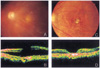
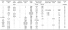
The mean postoperative follow-up time was 20.58 ± 7.24 months (range : 12 to 35). The postoperative results are shown in Table 1. In 13 of the 19 eyes, the macular hole was closed and the retina was reattached with one operation. The hole was closed with one operation in 15 eyes (79.0%). In six eyes, retinal detachment recurred during the postoperative follow up period due to reopened macular hole in four eyes and new or missed peripheral retinal break in two eyes. In one case of reopened macular hole (#8), fluid-gas exchange with none-expansile sulfur hexafluoride and subsequent laser photocoagulation on the edge of the macular hole was performed on an out-patient basis. In three cases of reopened macular hole (#4, 11, 16) with prominent posterior staphyloma, macular buckling was performed with silicone sponge (Mira type 506). After these procedures, the reopened macular hole was successfully closed and the retina was flattened. Encircling was applied in two cases with recurred retinal detachment with peripheral retinal breaks (#6, 7) to reattach the retina. In all 19 cases, the macular hole was sealed and the retina was reattached after including the second operation. Macular hole closure was confirmed not only by slit lamp examination with +90 D lens but also by optical coherence tomography (Fig. 1).
The best-corrected visual acuity was improved in 11 eyes (57.9%), unchanged in seven (36.8%), and deteriorated in one (5.3%). Preoperative mean visual acuity was 0.05 and postoperative mean visual acuity was 0.15. Cataracts developed in two eyes during the follow-up period and cataract surgery was performed.
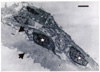
TEM photograph of the excised specimen revealed that ILM was present in all specimens (Fig. 2). Hyperplastic retinal pigment epithelial cells and/or fibrous astrocytes attached on the vitreous side of ILM were almost universally found.
DISCUSSION
The onset mechanism of retinal detachment caused by macular hole in the highly myopic eye is not fully understood.1 The theory that idiopathic senile macular hole is developed by traction caused by focal shrinkage of the vitreous cortex in the foveal area has been accepted so far.11 It could be assumed that the onset mechanism of macular hole in high myopia might be similar to that of idiopathic macular hole.12 However, some differences exist between the macular holes of high myopia and those of the senile idiopathic type. Development of retinal detachment is relatively common in myopic macular holes in contrast to idiopathic macular holes. Several factors have been postulated as contributing to the development of retinal detachment in myopic macular hole. The atrophy of the retinal pigment epithelium and choroids, posterior staphyloma, and tangential vitreous traction were thought to be important factors of this disease.2,3 The active transport of ions across the retinal pigment epithelium and oncotic pressure from the protein-rich choroidal fluid are the major forces which draw fluid out of the subretinal space.13 The atrophy of the retinal pigment epithelium and choroids would eventually weaken the adherence of the sensory retina to the retinal pigment epithelium. The myopic chorioretinal change may represent a spectrum evolving from tigroid to the widespread diffuse atrophy. The myopic eye with tigroid fundus provides a base conducive to the development of retinal detachment, and severe chorioretinal atrophy further increases the incidence.12
Akiba et al revealed that retinoschisis and retinal detachment often develop in highly myopic eyes without macular holes by optical coherence tomographic study.14 It could be thought that a macular hole evokes retinal detachment more easily, and adversely, that a macular hole may form as a result of preexisting retinoschisis or retinal detachment.15 They found that extensive retinal detachment developed in 95% of eyes with macular holes and myopia (over -6D) with a posterior staphyloma, compared to in only 7% of eyes without posterior staphyloma. They concluded that posterior staphyloma rather than anteroposterior vitreomacular traction may contribute to the development of retinal detachment associated with a macular hole in severely myopic eyes.16 The ectatic sclera in high myopia is associated with a number of abnormalities such as extreme thinning, absent interweaving of the fiber bundles, and their dissociation.17 The sclera of the posterior pole is stretched in the eyes with posterior staphyloma, resulting in a disproportion among sclera, choroid and retina. In this situation, due to "inverse traction", the sensory retina becomes easily detached from the pigment epithelium.4,12
Although the tangential and anteroposterior vitreous traction were thought to be eliminated through the removal of perifoveal ILM, redetachment via reopening of the myopic macular hole was still substantial incidence (21%) in our study. This may suggest the role of the posterior staphyloma.
Ishida et al investigated the ultrastructure of surgically removed epiretinal membrane.1 The epiretinal membrane consists of cortical vitreous and various cellular components. The active synthesis of new collagen aggregates regulated by fibrous astrocytes in a synchronized cellular network, with communication by gap junctions and active cell-matrix interactions, may lead to a diffusely condensed posterior cortical vitreous. This cell-mediated remodeling of the collagen matrix of the posterior cortical vitreous may generate tangential traction and play an important role in the pathogenesis of myopic macular hole with retinal detachment.1 Stirpe et al reported that tangential traction from the epiretinal tissue was the apparent cause of recurrent retinal detachment in highly myopic eyes with macular hole.18 In this study, TEM photographs similarly showed that fibrous astrocytes were adhered to removed ILM(Fig. 2).
In 2000, Gonvers, based on his experience of treatment for retinal detachment owing to myopic macular hole, reported that the choice of the surgical technique is best based on (1) the presence or absence of a posterior staphyloma and/or (2) chorioretinal degenerations and on (3) the axial length of the globe.19 A recent report by Ripandelli et al indicates that the vitreous anatomy such as posterior vitreous detachment or posterior vitreous schisis should be considered in deciding the surgical approach.20
The role of the posterior vitreous detachment in the development of retinal detachment in myopic eyes with macular hole is controversial. Morita et al suggested that posterior vitreous detachment does not act as substantial causative factor in producing retinal detachment from macular hole.12 On the other hand, Akiba et al reported that extensive retinal detachment developed in 89% of cases with complete posterior vitreous detachment and in 31% of cases without posterior vitreous detachment, and that the difference was significant.16
Many surgical techniques have been tried for the treatment of retinal detachment caused by macular hole in the highly myopic eye.4,5 Macular buckling and a method of retinopexy, with cryotherapy, diathermy, or photocoagulation macular diathermy, were used for treatment of the disease. The chief disadvantage of these techniques is the difficulty of approaching the macula due to the buckling and permanent destruction of the macular area.4 In 1982, Gonvers reported a new surgical technique for the treatment of retinal detachment due to myopic macular hole. This approach was based on the concept that vitreous traction plays a major role in the development of many of these detachments.4 In 1986, Miyake described a simplified method - subretinal fluid drainage and gas tamponade - of treating retinal detachment with macular hole. The advantages of this procedure are that it is easy, safe and requires no sophisticated instruments.10 Eyes with low myopia (<-10.0D) and without posterior staphyloma are best treated with vitrectomy and air/gas injection alone, whereas eyes with more marked (>-15.0D) and extensive atrophy and/or posterior staphyloma are best treated with vitrectomy, laser photocoagulation of the macular hole rim under perfluorocarbon liquid, and a temporary tamponade with silicone oil. Against the effect of posterior staphyloma, macular buckling has been tried. However, it was not easy to gain access to the macular area without vitrectomy, so a special exoplant and the surgical technique of posterior episcleral buckling were introduced.6,7 In our four cases, reopening of macular hole with retinal detachment was developed after initial surgery. In three of these cases, macular buckling was performed to release the reverse traction due to severe posterior staphyloma. In the other case, retinal reattachment was achieved by additional intravitreal gas injection followed by photocoagulation to the margin of the macular hole.
Tangential macular traction may play an important role in macular hole formation and subsequent retinal detachment.3,21 In this regard, it is important to perform epiretinal membrane separation as completely as possible.3,21 Recently, Kadonosono et al revealed the contraction of myofibroblasts on the ILM surface around the macular hole in highly myopic eyes with macular hole.2 This suggests that ILM surrounding the myopic macular hole plays a role in reopening of the hole and subsequent development of retinal detachment. The removal of ILM and overlying epiretinal membrane may increase the flexibility of the detached retina and it may contribute to sealing of the macular hole even in the presence of posterior staphyloma.2 Primary removal of ILM of the detached retina may also allow complete removal of the overlying epiretinal membrane.
Overall initial reattachment rates of 60% to 91% have been reported for the treatment of myopic retinal detachment caused by a macular hole in the previous literature.2,3,5 In 1990, Kuriyama et al evaluated reattachment rates of the transscleral and transvitreal techniques for treating retinal detachment due to macular hole and reported that the transscleral approach was superior in initial success rate (83% vs. 56%).5 Other initial success rates that have been reported include vitrectomy with or without an adjuvant agent such as transforming growth factor-β2 or platelet extract (60.0%),22 vitrectomy with complete epiretinal membrane separation (64.7%),3 vitrectomy with fluid-gas exchange and endolaser around the macular hole (62.5%),23 simplified gas injection method (83%)10, and vitrectomy with ILM removal (70~91%).2, 24 The previous reports indicated that the incidence of recurrent retinal detachment increases with a longer follow-up period. Although the initial reattachment was hindered due to the postoperative development of peripheral retinal breaks in a few cases, the initial macular hole closure rate of 79.0% is encouraging. However, the closure of the macular hole is sometimes difficult to attain, despite the complete relief of traction by the removal of ILM. This result suggests that the imbalance between the retina and the choroid-sclera complex associated with axial elongation and posterior staphyloma might be involved in the development of retinal detachment by myopic macular hole.
Overall, the initial success rate of myopic macular hole closure after vitrectomy with ILM removal seems higher than that of the previous studies. Moreover, this technique eliminates the necessity of a second vitreous surgical procedure, even in the case of recurred retinal detachment. We think that perifoveal ILM may be an important adjuvant to treat the myopic macular hole with retinal detachment.
 XML Download