Abstract
Intraspinal bronchogenic cysts are rare congenital cystic lesions. In all the reported cases, the cysts have been located in the cervical, upper thoracic or thoracolumbar segments. We report the case of an intraspinal bronchogenic cyst in the sacral location. We present the case of a 5-month-old female with a skin dimple in the midline over the sacral vertebra. Magnetic resonance image of the lumbar and sacral vertebra revealed a dermal sinus tract and an epidural cystic mass at the S2 level. The patient underwent the removal of the dermal sinus tract and the cyst. The cystic mass was shown to be connected to the subarachnoid space through a slender pedicle from the dura. The cyst was diagnosed to be a bronchogenic cyst based on the results of the histopathological examination. We conclude that intraspinal bronchogenic cysts may appear in the sacral location.
Endodermal cysts are presumed to be derived from the endoderm of the developing gastrointestinal tract or, in rare cases, the respiratory system. If the endodermal cyst is predominantly lined with respiratory tract epithelium, it is termed a bronchogenic cyst. Intraspinal bronchogenic cysts are rare, and their site of predilection is the cervical or upper thoracic segments generally with an intradural extramedullary localization. We report a rare case of an intraspinal bronchogenic cyst localized to the sacral vertebra and associated with other malformations, which was surgically treated.
A 5-month-old female was admitted to our Neurosurgery department with a skin dimple in the sacral area. On admission to hospital, the patients general examination revealed no abnormalities except for a small skin dimple in the midline over her sacral vertebra. Magnetic resonance imaging (MRI) of the lumbar spine and sacrum was conducted at another hospital and revealed a 1.3×0.5 cm oval lesion at the S2 level. The lesion was homogenous and hyperintense on T2-weighted images and hypointense on T1-weighted images. Further, MRI revealed a dermal sinus tract at the S2 level and a tethered spinal cord (Fig. 1).
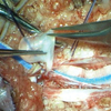
The surgery was performed under general anesthesia in the prone position. A midline cutaneous incision was made and the sinus tract was dissected out from the subcutaneous tissues. The presence of spina bifida was observed. The attachment of the tract to the dura was excised in an elliptic manner, and the dura was opened to visualize the subdural and the subarachnoid spaces immediately under the fibrous tract. The cystic mass was connected to the subarachnoid space through a slender pedicle from the dura (Fig. 2). Puncture of the mass yielded clear and colorless liquid, which was completely removed. The dura mater was then closed. There were no complications during the surgery. After 9 days, she underwent an MRI of the lumbar spine that revealed the complete excision of the dermal sinus and cyst and preservation of the neural tissues.
Histopathological examination revealed a bronchogenic cyst and a dermal sinus tract. The cyst was lined by one layer of ciliated columnar epithelial cells, and connective tissue was observed in the cystic wall. There were no mucus glands or cartilage (Fig. 3). These pathological findings were concordant with those of endodermal cyst with respiratory epithelium; therefore, we classified it as a bronchogenic cyst.
Endodermal cysts have been described using various names, including enteric cysts, enterogenous cysts (1), neurenteric cysts (2), gastrocytomas (3), teratomatous cysts (4), and archenteric cysts (5). They are presumed to be derived from the endoderm of the developing gastrointestinal tract or, in rare cases, the respiratory system. These are termed neurenteric and bronchogenic cysts, respectively. Endodermal cysts are typically intradural extramedullary lesions (6), most commonly located at the level of the cervical spine (6, 7); moreover, a ventral location is common (8). Other malformations of the spine are commonly associated with this disease entity. such as fused vertebrae, spina bifida, and hemivertebrae. The histological examination of the endodermal cyst wall typically shows a mucus-secreting columnar cell lining. In some instances, cilia may be demonstrated (7, 9). On light microscopy, the bronchogenic variant can be differentiated by the presence of ciliated and nonciliated cells and goblets cells of the normal tracheobronchial tract. The cyst's wall may contain mucus and serous glands, cartilage, smooth muscle, and ganglia (10). In our patient, light microscopy revealed the bronchogenic variant.
Bronchogenic cysts are found most frequently along the tracheobronchial tree in the mediastinum or with the lung parenchyma. Rarely, these cysts have occur in other locations, including cutaneous (11) and subcutaneous tissues (12), the pericardium (13), the diaphragm (14), the abdomen (15), and the spinal cord. To our knowledge, six intradural extramedullary bronchogenic cysts have been described in the literature, further, all of them were localized in the cervical, upper thoracic, or thoracolumbar segments (10, 16-20). Thus, we report the first sacral intraspinal bronchogenic cyst.
The main hypothesis regarding the development of these malformations propose a faulty separation between the ectodermal and endodermal layers resulting in the inclusion of endodermal tissue within the ectodermally derived spinal cord (21). Incomplete excalation of the chordal plate would lead to the persistence of the neurenteric canal and the associated anomalies of the notochord would explain the commonly associated spinal and chordal anomalies (22, 23). Due to the further differentiation of the endodermal layer into the digestive or respiratory tract, endodermal cysts may present with both types of histological features.
In conclusion, intraspinal bronchogenic cysts are uncommon congenital malformations. Although these cysts are generally located in the cervical, thoracic, or thoracolumbar spine, a sacral location should also be considered. Moreover, in such cases, the early recognition of associated vertebral or spinal cord anomalies is essential.
Figures and Tables
References
2. Holcomb GW Jr, Matson DD. Thoracic neurenteric cyst. Surgery. 1954. 35:115–121.
3. Knight G, Griffiths T, Williams I. Gastrocystoma of the spinal cord. Br J Surg. 1955. 42:635–638.

4. Hoefnagel D, Benirschke K, Duarte J. Teratomatous cysts within the vertebral canal: observations on the occurrence of sex chromatin. J Neurol Neurosurg Psychiatry. 1962. 25:159–164.

5. Neuhauser EB, Harris GB, Berrett A. Roentgenographic features of neurenteric cysts. Am J Roentgenol Radium Ther Nucl Med. 1958. 79:235–240.
6. Eynon-Lewis NJ, Kitchen N, Scaravilli F, Brookes GB. Neurenteric cyst of the cerebellopontine angle: case report. Neurosurgery. 1998. 42:655–658.
7. Miyagi K, Mukawa J, Mekaru S, Ishikawa Y, Kinjo T, Nakasone S. Enterogenous cyst in the cervical spinal canal. Case report. J Neurosurg. 1988. 68:292–296.
8. Macdonald RL, Schwartz ML, Lewis AJ. Neurenteric cyst located dorsal to the cervical spine: case report. Neurosurgery. 1991. 28:583–587.

9. Morita Y, Kinoshita K, Wakisaka S, Makihara S. Fine surface structure of an intraspinal neurenteric cyst: a scanning and transmission electron microscopy study. Neurosurgery. 1990. 27:829–833.

10. Ho KL, Tiel R. Intraspinal bronchogenic cyst: ultrastructural study of the lining epithelium. Acta Neuropathol. 1989. 78:513–520.

11. Tresser NJ, Dahms B, Berner JJ. Cutaneous bronchogenic cyst of the back: a case report and review of the literature. Pediatr Pathol. 1994. 14:207–212.

14. Buddington WT. Intradiaphragmatic cyst: ninth reported case. N Engl J Med. 1957. 257:613–615.
15. Coselli MP, Ipolyi P, Bloss RS, Diaz RF, Fitzgerald JB. Bronchogenic cysts above and below the diaphragm: report of eight cases. Ann Thorac Surg. 1987. 44:491–494.

16. Baba H, Okumura Y, Ando M, Morioka K, Noriki S. A high cervical intradural extramedullary bronchogenic cyst. Case report. Paraplegia. 1995. 33:228–232.

17. Baumann CR, Könü D, Glatzel M, Siegel AM. Thoracolumbar intradural extramedullary bronchogenic cyst. Acta Neurochir (Wien). 2005. 147:317–319.
18. Rao GP, Bhaskar G, Reddy PK. Cervical intradural extramedullary bronchogenic cyst. Neurol India. 1999. 47:79–81.
19. Wilkinson N, Reid H, Hughes D. Intradural bronchogenic cysts. J Clin Pathol. 1992. 45:1032–1033.

20. Yamashita J, Maloney AF, Harris P. Intradural spinal bronchiogenic cyst. Case report. J Neurosurg. 1973. 39:240–245.
21. Cheng JS, Cusick JF, Ho KC, Ulmer JL. Lateral supratentorial endodermal cyst: case report and review of literature. Neurosurgery. 2002. 51:493–499.

22. Korinth MC, Muller HD, Gilsbach JM. Neurenteric cyst of the cervical spine with mediastinal extension: case illustration. J Neurosurg. 2003. 98:1 Suppl. 112.


 PDF
PDF ePub
ePub Citation
 Print
Print


 XML Download