

 PDF
PDF ePub
ePub Citation
 Print
Print


INTRODUCTION
Brucellosis is a zoonosis that has a worldwide distribution, and it remains as a major source of disease for both humans and domestic animals particularly in the Mediterranean region, Western Asia and in parts of Africa and Latin America (1). According to a review of the literature, bovine brucellosis was eradicated in Japan in 1992 and in North Korea in 1959 (1). Although human brucellosis has been rarely reported in the East Asian countries until now, bovine brucellosis has been sporadically reported since 1947 and it has increased to above 100 cases per year since 1984 in the Republic of Korea (ROK) (2, 3). After 1990, the cases of bovine brucellosis have increased to above 500 cases every year (3-6). Furthermore, in 2003, 1,088 cases of bovine brucellosis were reported in ROK (7). Because of increasing trend for bovine brucellosis in ROK, there is now a greater possibility for humans to become infected with brucellosis than at any time during the past 2 decades. Although several studies have been performed to detect human brucellosis, they did not find any such cases (4-6). The first suspected case of human brucellosis in ROK was that of a livestock worker in 2002 (3). Recently, there was a report of an outbreak of human brucellosis among livestock workers and veterinarians in rural area around Jeongeup City, Jeollabuk-Do, Korea, from February 2003 to August 2003. Although brucellosis has mainly been a problem of veterinarians for past five decades, this local sporadic outbreak demonstrated that Brucella abortus biotype 1 in humans has emerged as an important public health problem in ROK.
In this report, we describe the clinical characteristics and the serologic data in the II cases of human brucellosis and also discuss and the efficacy and side effects of a rifampin and doxycyline regimen for the treatment of human brucellosis.
MATERIALS AND METHODS
Patients
An epidemiological investigation was conducted to identify the vehicle and the source of the brucellosis infection. We also tried to describe the circumstances of the outbreak and establish the control measures to be taken by the Division of Zoonoses, Center for Immunology and Pathology, National Institute of Health, Korea.
Jeoungeup City is an administrative district region consisting of Sintaein Eup, fourteen Myens including Ipyong, Deokcheon, and Gobu, and 15 Dongs. These regions are very close to northwest Jeongeup City. In 2003, the cow stock in Sintaein, Ipyong, Deokcheon and Gobu accounted for 16.8% of the total cow population of the Jeongeup City region.
A suspicious case of human brucellosis was defined as a person who was residing in regions of Sintaein, Ipyong, Deokcheon or Gobu, and who presented with vague clinical symptoms of brucellosis such as fever of unknown origin, unexplained weight loss, fatigue etc. from February 2003 to August 2003. A serologic screening with the standard tube agglutination test (STA) was done in fifty patients who had a history of a contact with cows diagnosed with bovine brucellosis and who also had the unexplained vague symptoms described above. The eleven probable cases among the fifty suspicious cases included patients who had a positive STA titer of ≥1:160, a positive PCR result, or positive scrological results by ELISA. A confirmed case was defined as a serologically diagnosed case that revealed a positive blood culture (8).
Serological and microbiological culture methods
The serologic tests were performed regularly starting at the patients' first visit to the district health service center; the tests were repeated six weeks after the start of treatment and at 8, 12, 16, 20, 24 (at 4 weeks, interval for 6 months) and 48 weeks later.
The district health service center referred the blood samples to the Division of Zoonoses, Center for Immunology and Pathology, National Institute of Health, Seoul, Korea. The blood cultures were processed with the automatic blood culture system (BACTEC 9050, BD Co, Sparks, Maryland, U.S.A.), and they were incubated for 5 to 7 days. If there were any positive signs, a "blind" subculture was done with Tryptic Soy agar (containing 5% sheep blood, Difco, Detroit, MI, U.S.A.) for 2 to 3 days in 5% CO2 at 37℃. The human isolates of the Brucella strain were identified using the standard method (9).
STA and ELISA for the IgM and IgG antibodies to B. abortus were performed for each serum sample. All the samples from the same patient were processed simultaneously by the progressive double-dilution method. Different antigens were also used for detection in each of the assays. A suspension of B. abortus antigen (Difco Laboratories) was prepared as the antigen for the tube agglutination test. A B. abortus diagnostic kit (Pan Bio, Brisbane, Australia) was used for the ELISA IgG and IgM tests. To find any cross-reactivity with other Gram-negative bacteria, we performed STA procedures with Yersinia enterocolitica (ATCC 9610), and Francisella tularensis (Germaine, San Antonio, Texas, U.S.A.). However, no cross-reactivity was noted. Although there is no single titer of Brucella antibodies that is 100% diagnostic, most cases of active infection have titers of 1:160 or greater for IgM, along with positive IgG antibodies. The cut-off values were 11 for the IgM and IgG.
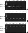
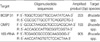
The template DNA was extracted by using a blood DNA purification kit (Gentra, Minneapolis, Minnesota, U.S.A.) from the blood of the patients. PCR was performed in a 50 µL volume containing the following; 10 µL template DNA, 0.025 U Taq DNA polymerase (Promega, Madison, WI, U.S.A.), 3 µL MgCl2 (25 mM), 5 µL 10×PCR reaction buffer (1.5 mM), 1 µL PCR Nucleotide Mix (10 mM each), and 10 pM of each primer. The primers were genus-specific primer pairs designed to amplify the gene encoding a 31-kDa, 36-kDa and 16S rRNA fragment of the genus Brucella (9, 10) (Table 1). The cycling condition included an initial denaturation step at 94℃ for 10 min, and the template was cycled 35 times (1 min of denaturation at 95℃, 1 min of annealing at 60℃ and 5 min of extension at 72℃) by a thermal cycler (Perkin-Elmer PCR system 9700). The positive control contained B. abortus ATCC 7705 DNA (biotype 1) as the template, and the negative control consisted of sterile water instead of the DNA template. The PCR products were resolved by electrophoresis on 2% agarose gel with ethidium bromide (0.5 mg/mL).
RESULTS
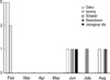
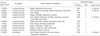
A total of eleven cases were identified at the local outbreak; the cultures were positive in four cases, and seven cases were serologically diagnosed (Table 2). Fig. 1 shows the distribution of the patients who presented with human brucellosis in the Jeougeup city area, Jeollabuk-Do, Korea. Eight patients were male and 3 patients were female, and their ages ranged from 38 to 51 yr old (mean, 45 yr). Ten patients were livestock workers and one patient was a veterinarian who acquired the disease through an accidental contact with infected cows while assisting in calf delivery. In our study, all the cases were interviewed to discover the mode of transmission. However, they denied buying or consuming unpasteurized milk, but they did consume raw meat, including raw muscle and liver. Case 3, 8 and 9 had a history of eating uncooked fresh muscle and liver. Although we could not get information about how many cows were infected or concerning their level of disease because the cattle had already been slaughtered, the confirmed Brucella biotype 1 bacteria strongly suggested that respiratory transmission or direct contact transmission was responsible for the human infections rather than ingestion of the cattle meat or milk. The patients were probably infected by their occupational and incidental contact with infected cattle.
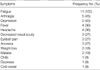
The most frequent symptom was fatigue (100%), and other common complaints are listed in Table 3. The ophthalmologic examinations revealed only senile cataract changes without any definitive pathognomonic findings.
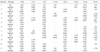
Table 4 shows the one year follow-up serologic data for one year. The median STA antibody titer for the eleven patients at the time of their admission was 1:160 (range, 1:40-1:640). The mean IgM ELISA titer was 57.5 (range, 16-173), and the mean IgG ELISA titer was 35 (range, 11-76). We used 3 different kinds of primer sets to perform PCR, and all of them showed positive results (Fig. 2). There were 4 positive blood cultures (36%) at the time of admission. The initial STA titers of cases 2, 7 and 9 were below the diagnostic value, but their follow-up titers increased four fold after several weeks and the results of their PCR tests were all positive. Especially, case 2 had a positive blood culture result. The IgM antibody test results obtained from the sera at the time of admission were all above the cutoff levels in all patients, but the IgG levels remained within the normal range in 4 cases (case 3, 4, 7 and 9) (Table 4). Following treatment, the IgM antibody levels decreased steadily; however, the IgG levels increased at first, but they steadily decreased thereafter.
The patients' treatment was started based on the positive serologic findings in the suspicious cases because the disease symptoms were nonspecific. The treatment regimen consisted of oral doxycycline 100 mg twice a day plus oral rifampin 900 mg/day in a single morning dose, and this regimen was given from 8 weeks to a maximum of 10 weeks. Among the 11 patients, one patient had a relapse 5 months after the completion of the therapy. In this case, the clinical symptoms of severe fatigue, anorexia and weight loss recurred; the blood culture was positive and the serologic titers for the IgM and IgG by ELISA were sharply increased. In addition, the titer of the STA was increased over four fold. This relapsed patient was re-treated with the same antibiotic regimen for additional 6 more weeks, and then his symptoms and serologic titers improved. We could not find any definitive reason for the relapse. However, we thought that poor compliance with taking his medication rather than drug resistance might have been the primary problem, or reinfection was also a possibility. The treatment regimen was relatively well tolerated, but gastrointestinal disturbance was the major complaint from the medication. One patient showed melena and hematemesis after completion of the medication schedule. She continuously complained of gastrointestinal problems and pain while taking the medication, but she completed the drug course. One week after the conpletion of the medication schedule, she was diagnosed with hemorrhagic esophagitis by flexible gastrofiberscopy.
There had been no previous cases of human brucellosis in these areas. The first series of cases presented with symptoms on February 2003 and the last case presented on August 2003. As a consequence, the outbreak duration was deemed to be about 7 months long.
DISCUSSION
Because brucellosis has a wide spectrum of nonspecific clinical manifestations, this disease may clinically mimic another febrile illnesses such as typhoid fever and tuberculosis. In addition, healthcare providers may not consider brucellosis in their first differential diagnosis. Therefore, there is a strong possibility that the disease may be considerably underdiagnosed.
The possibility of human beings falling ill with brucellosis has been a reality for a long time in the ROK because bovine brucellosis has been continuously reported on for many decades. After 1990, the number of cases of bovine brucellosis has increased to above 400 every year (3-6). Chung et al. reported that the major B. abortus was biotype 1 in Korean dairy cattle (11). In this study, the species of Brucella identified was also biotype 1. Therefore, this microbiological result strongly suggests that B. abortus biotype 1 might be a major pathogenic species in ROK. Infected cattle shed this organism into the environment via urine, vaginal secretions, ejaculates, aborted fetuses or feces. Baek et al. insisted that an indigenous Korean dog was infected via contacts with infected cattle on the same farm where the dog was living (12).
In 2003, there were more than 1,088 cases of bovine brucellosis; about 60% of the bovine brucellosis cases in 2003 occurred in the Korean native cattle and the other cases were in the dairy cattle (6). Korean native cattle are raised to produce meat, not for milk. By contrast, human brucellosis in the Mediterranean regions is acquired from the dairy cattle, goats, camels and sheep, and these animals can produce contaminated milk and cheese. So, there is a big difference in the cause of human brucellosis between ROK and the Mediterranean area. It was thought that brucellosis in ROK might be transmitted through abraded skin that incurred during the course of handling infected animals or their carcasses rather than through the ingestion of unpasteurized dairy products. This epidemiological difference suggests that anti-epidemic measure for brucellosis in ROK should be focused on the control of the Korean native cattle as well as the dairy cattle.
The symptoms in Korean patients were somewhat different from twose in patients from other countries. Fever, chills and sweating were the common symptoms, and these were also the symptoms in more than 70% of the cases from other countries. Arthralgia and cough were also common in the cases from other countries (13). Of note, fatigue was the most common symptom, occurring in all patients.
The culture techniques are time-consuming and they lack sensitivity for patients having chronic infections; furthermore, handling these organism in the laboratory is hazardous (14, 15). PCR is a useful modality for the rapid and direct detection of Brucella DNA in the blood specimens obtained from persons with brucellosis, and it can provide results to the clinician in less than 24 hr. Moreover, it eliminates the hazards of handling this organism in the laboratory (16-18). In our study, PCR was very sensitive. It displayed 100% positive results. Although blood culture has been held as the gold standard for the laboratory diagnosis of brucellosis until now, the sensitivity of this technique is very low, ranging from 15% to 70% (19). Therefore, PCR should also be performed in hospitals, especially in the endemic countries.
In our study, the IgM antibody levels determined by ELISA on the sera obtained at admission were all above the cutoff levels in all patients, but the IgG levels remained within the normal range in 4 cases. Irmak et al. (20) reported that although the majority of the patients may present with specific IgM antibodies as well as with specific IgG antibodie, other cases may have either specific IgM or specific IgG antibodies. So in clinical practice, the two assays should be performed as complementary tests.
During the follow-up period, case 1 again had positive blood cultures; this patient experienced a relapse of the disease about 5 months after the end of treatment. In this case, the IgM/IgG antibodies levels were markedly and sharply elevated. Although the IgM/IgG antibody levels were markedly elevated in case 3, the patient had no symptoms and the blood cultures were negative. Several reports have demonstrated that patients in relapse often show an elevation of the IgG level by ELISA (9, 21). By contrast, as was reported by Gazapo et al. (10), the IgM level by ELISA did not elevate. Yet Irmak et al. (20) have shown that the IgM/IgG antibodies levels were elevated in relapsed cases, in line with our observation but the IgM titers were not markedly increased compared with the IgG titers.
The treatment regimen in this study consisted of oral administration of doxycycline 100 mg twice a day plus oral rifampin 900 mg/day in a single morning dose for 8 weeks to a maximum of 10 weeks. There was only a single case of relapse (9.1%). Solera et al. (22) have strongly suggested that the doxycycline/rifampin regimen is less effective than the doxycycline/streptomycin regimen in patients with acute brucellosis. The relapse rate in the doxycycline/rifampin group was 16%, white it was only 5.3% in the doxycycline/streptomycin group. Montejo et al. (23) have reported that the relapse rate in the doxycycline/rifampin group was 10.8%. There was only one relapse case in our study; however, we could not differentiate between relapse and reinfection with certainty. The treatment duration in our study was longer than in other studies because some patients wanted to have longer medication; the treatment duration in other studies was around 6 weeks (42-45 days) with the doxycycline/rifampin regimen (21-23).
In conclusion, this outbreak shows that the ROK is no longer free of human brucellosis. Those livestock workers who are suffering with chronic fatigue, arthralgia, depression, fever and headache without any apparent reasons should visit a physician for a medical evaluation. The physicians who are living and working in the regions with endemic bovine brucellosis have to be aware of the clinical manifestations of human brucellosis and the necessary workup to establish the diagnosis when indicated.
 XML Download