

 PDF
PDF ePub
ePub Citation
 Print
Print


Introduction
Compared with the transfemoral approach, transradial coronary intervention is valuable in terms of major vascular access related complications and early mobilization.1)2) However, this approach has a major limitation in its use of the large (7 or 8 Fr sheaths) bore-guiding catheter with the relatively small size of the radial artery,3)4) as such may result in a reduced procedural success rate as well as an increased incidence of post-procedural radial artery occlusion.5) The sheathless guide catheter system (Sheathless Eaucath, Asahi Intecc®, Nagoya, Japan) is a newly developed device, composed of a hydrophilic catheter and a long central dilator. This dedicated system enables us to introduce the catheter into the artery without an introducer sheath, which can decrease the outer diameter of guiding catheter system and provide less invasive angioplasty and a puncture site injury compared with a conventional guiding catheter system with introducer sheath. However, these devices are not currently available in Korea. We present our experience in which a sheathless transradial coronary intervention was performed using a standard guiding catheter instead of a dedicated sheathless guide catheter system.
Case
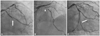
The patient was an eighty year-old male, experiencing exertional chest pain for 3 months. His coronary risk factors were hypertension and smoking. The electrocardiography and cardiac enzymes were unremarkable. The echocardiography showed normal left ventricular systolic function without regional wall motion abnormalities. The coronary angiography via the right radial artery demonstrated total occlusion at distal left circumflex artery (LCX), and diffuse 70-90% stenosis with severe calcification from distal left main coronary artery to proximal left anterior descending artery (LAD) (Fig. 1A and B). The right coronary angiography showed no significant stenosis. We planned to treat the distal LCX occlusion first. A 6 Fr EBU 3.75 guiding catheter (Medtronic®, Minneapolis, MN, USA) was engaged to the left coronary artery (LCA) by a conventional transradial technique. A Fielder FC wire (Asahi Intecc, Nagoya, Japan) successfully crossed the occlusion, and a Xience prime 2.5×33 mm stent (Abbott®, Redwood City, CA, USA) was implanted at distal LCX across the second obtuse marginal branch (OM) after several predilatations. Following a good stent implantation distal LCX flow, however, a dissection occurred at second OM, perhaps needing another stent implantation through the stent strut (Fig. 1C). As the patient did not complain of chest pains and the vital signs were stable, we decided on the staged percutaneous coronary intervention (PCI) for the distal LCX bifurcation stenting and the severe calcified proximal LAD stenosis with a larger bore guiding catheter.
Two days after the procedure, a second PCI was performed via a radial artery, as the patient was not able to lie straightly due to right leg pain. Because a large bore guiding catheter system was required for this complex coronary anatomy, we decided to perform the sheathless transradial approach using a standard 7 Fr guiding catheter. At first, a 5 Fr introducer sheath was inserted into the right radial artery. Then a stiff 260 cm J-tipped 0.035-inch wire (Terumo®, Tokyo, Japan) was introduced near the aortic root. After the removal of the 5 Fr introducer sheath, we predilated radial artery access site with a dilator of the 7 Fr introducer sheath.
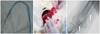
Following, we inserted the Heartrail catheter (Terumo®, Tokyo, Japan) into the 7 Fr XB 3.5 guiding catheter (Cordis®, Miami, FL, USA) (Fig. 2A). Both catheters were inserted sequentially through the skin into the radial artery (Fig. 2B), and advanced to the aortic root along the 0.035-inch wire (Fig. 2C). Finally, 7 Fr XB 3.5 guiding catheter was successfully engaged into the LCA.
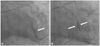
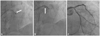
The first target lesion was the distal LCX bifurcation lesion. After a guide wire was passed into the second OM, multiple predilatations were performed, and Xience prime 2.5×23 mm stent was deployed by culottes technique across the previous LCX stent (Fig. 3A). Thereafter, kissing balloon dilatation was successfully performed (Fig. 3B). Because proximal LAD target lesion had a severe calcification, rotational atherectomy (1.5 mm burr) was performed five times (Fig. 4A), and 2 Xience prime stents (2.75×15 mm, 3.5×23 mm) were implanted with the support of 5 Fr, 120 cm Heartrail catheter (child-in-mother technique) after multiple predilatations (Fig. 4B).
The final angiography revealed excellent results (Fig. 4C). The patient was stable without elevation of enzymes, and was discharged without puncture site complication two days after the procedure.
Discussion
Several studies have begun to outline the advantages, safety, and efficacy of transradial procedures in both low- and high-risk patient populations.4)6) Unfortunately, the size of guiding catheters used in transradial coronary intervention is limited by anatomical considerations. The lumen of the radial artery is frequently smaller than the outer diameter of a 7 or 8 Fr radial sheath. Dahm et al.5) suggests that the over-sized sheaths in relation to the size of the radial artery may contribute to acute arterial occlusions. Although asymptomatic and rarely associated with ischemia, radial artery occlusions sometimes prohibit subsequent radial artery access.7)
As we have shown in this case, to avoid such potential radial complications in complex transradial coronary intervention, sheathless guiding catheter can be used. The outer diameter of the 7 Fr XB 3.5 guiding catheter (2.31 mm) is smaller than the outer diameter of the 6 Fr introducer sheath (2.52 mm). Thus, sheathless guiding catheter insertion allows complex procedures requiring the internal lumen of a 7 Fr guiding catheter, such as, rotational atherectomy, culottes technique, kissing balloon technique, and crushing stent technique. The sheathless guide catheter system has recently been introduced. The sheathless guide catheter system is composed of a hydrophilic guiding catheter and a long central dilator. However, these devices are currently not available in Korea. Thus we performed sheathless transradial coronary intervention with a standard 7 Fr guiding catheter and a 5 Fr Heartrail catheter.
We used a stiff J-tipped guide wire for better support and predilated the radial artery with a dilator of the 7 Fr introducer sheath to overcome diameter gap between radial artery access site and a 5 Fr Heartrail catheter tip. A "pseudo-taper" was created by insertion of a 5 Fr Heartrail catheter into and through the 7 Fr XB 3.5 guiding catheter. Thus, we could easily insert the 7 Fr guiding catheter through the skin into the radial artery by using a 5 Fr Heartrail catheter as a long dilator.
Furthermore, this technique for insertion of nonhydrophilic-coated guiding catheter may reduce the potential complications associated with long hydrophilic coating, such as granulomatous inflammation of the skin at the insertion site.8)9)
Three studies have reported successful use of a sheathless guiding catheter in complex interventions with large bore guiding catheters, including rotational atherectomy, crushing stent technique, intervention of bifurcation lesion, aspiration thrombectomy, simultaneous kissing stent technique, and modified T-stenting technique.10-12) These results suggests that other complex procedures may also be performed with the sheathless guiding catheter technique.
The learning curve of the sheathless guiding catheter technique may be steeper for inexperienced radial operators than the conventional guiding catheter systems. However, the sheathless guiding catheter technique is feasible in most clinical settings regardless of the complexity or severity of the lesions and associated with high rates of procedural success.
 XML Download