

 PDF
PDF ePub
ePub Citation
 Print
Print


Abstract
Purpose
Expression patterns, associated anomalies and progress of the patients with Goldenhar syndrome from the neonatal period were systematically investigated. This allows us to evaluate the need for early diagnosis.
Methods
This is a retrospective study of 29 infants with Goldenhar syndrome whose diagnosed in Samsung Medical Center between 1994 and 2013. Associated anomalies and procedures between neonatal group (n=13) and non-neonatal group (n=16) were systematically compared.
Results
Mean gestational age in the neonatal group were 38+1±2+4 weeks and 3 patients (23%) were preterm infants. The average birth weight in the neonatal group were 2,853±544 grams. Goldenhar syndrome was mainly diagnosed by ear and face anomalies during the neonatal period. The associated anomalies in neonatal group were cardiovascular anomaly (54%), genitourinary anomaly (30%), vertebral anomaly (15%), and others (31 %). About 40% of patients who had long-term follow-up revealed hearing abnormalities and about 1/4 of all patients had bilateral hearing problem, which resulted in requiring hearing aid devices. In addition, the most common procedure performed during follow-up was preauricular skin tag removal. And other procedures or surgery related to oral, eyes, and others were performed in each 1/4 of the patients. Cardiac surgery was done in 15% of total patients. Frequency of associated anomalies and performed procedures between the patients diagnosed at neonatal and non-neonatal period was not significantly different.
REFERENCES
1.Jones KL. Smiths recognizable patterns of human malformations. 5th ed.Philadelphia: WB Saunders Co;1997. p. 642–5.
2.Yun AY., Baek NS., Lee YA., Moon HR. Two cases of Goldenhar syndrome. J Korean Pediatr Soc. 1990. 33:380–5.
3.Song MY., Kim MS., Park NS., Hyoung UJ., Lee JO., Kim ER. Two cases of Goldenhar syndrome. J Korean Pediatr Soc. 1991. 34:730–5.
4.Hyun JE., Park EH., Jeon HY., Byeun WJ., Hwang YM. A case of Goldenhar's syndrome. J Korean Pediatr Soc. 1992. 35:135–9.
5.Ahn GH., Wee YS., Lee KH. A Case of Goldenhar's syndrome with vesicoureteral reflux. Korean J Perinatol. 2007. 18:303–7.
6.Grorlin RJ., Pindborg JJ. Oculoauriculovertebral dysplasia. In: Syndromes of the head and neck. 1st ed. New York: Mc-GrawHill Co, 1964: p. 419–26.
7.Goldenhaar M. Associations malformations de I' oeil et de I' oreille en particular Ie syndrome dermoide epibulbaire appendices auricularies - fistula auris congenita et ses relations avec la dysostose mandibulofaciale. J Genet Hum. 1952. 1:243–82.
8.Gorlin RJ., Jue KL., Jacobsen U., Goldschmidt E. Oculoauri-culovertebral dysplasia. J Pediatr. 1963. 63:991–9.

9.Feingold M., Baum JL. Goldenhar's syndrome. Am J Dis Child. 1978. 132:136–8.
10.Ballesta-Martínez MJ., López-González V., Dulcet LA., Ro-dríguez-Santiago B., Garcia-Miñaúr S., Guillen-Navarro E. Autosomal dominant oculoauriculovertebral spectrum and 14q23.1 microduplication. Am J Med Genet A. 2013. 161A:2030–5.
11.Strömland K., Miller M., Sjögreen L., Johansson M., Joelsson BM., Billstedt E, et al. Oculo-auriculo-vertebral spectrum: associated anomalies, functional deficits and possible developmental risk factors. Am J Med Genet A. 2007. 143A:1317–25.
12.Tasse C., Böhringer S., Fischer S., Lüdecke HJ., Albrecht B., Horn D, et al. Oculo-auriculo-vertebral spectrum (OAVS): clinical evaluation and severity scoring of 53 patients and proposal for a new classification. Eur J Med Genet. 2005. 48:397–411.
13.Baum JL., Feingold M. Ocular aspect of Goldenhar's syndrome. Am J Ophthalmol. 1973. 75:250–7.
14.Cohen MS., Samango-Sprouse CA., Stern HJ., Custer DA., Vaught DR., Saal HM, et al. Neurodevelopmental profile of infants and toddlers with oculo-auriculo-vertebral spectrum and the correlation of prognosis with physical findings. Am J Med Genet. 1995. 60:535–40.
15.Bogusiak K., Arkuszewski P., Skorek-Stachnik K., Kozakiewicz M. Treatment strategy in Goldenhar syndrome. J Craniofac Surg. 2014. 25:177–83.
16.Pashayan HI., Pinsky L., Fraser FC. Hemificial microsomia-oculoauriculovertebral dysplasia, A patient with overlapping features. J Med Genet. 1970. 7:185–8.
Fig. 1.
A Goldenhar syndrome patient was diagnosed at neonatal period with prominent and multiple preauricular skin tags (arrows) (A), facial asymmetry with right facial hypoplasia (B). He was followed-up after preauricular skin tag removal in plastic surgery at 2 month of age (C).
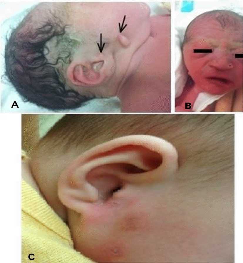
Table 1.
Prenatal and Postnatal Characteristics of the Enrolled Cases Diagnosed in the Neonatal Period
Table 2.
Comparison of Associated Anomalies between Enrolled Patients Diagnosed in the Neonatal Period (n=13) and Patients Diagnosed in the Non-neonatal Period (n=16)
| Systemic anomalies | Neonatal (n=13) | Non-neonatal (n=16) |
|---|---|---|
| Face | 6 (46%) | 7 (44%) |
| Hypoplasia of malar, maxillary or mandibular region | 3 (23%) | 4 (25%) |
| Asymmetry | 3 (23%) | 6 (38%) |
| Ear* | 13 (100%) | 11 (69%) |
| Microtia and dysmorphy | 7 (54%) | 8 (50%) |
| Tag and pit* | 9 (69%) | 4 (25%) |
| Inner anomaly | 2 (15%) | 1 (6%) |
| Oral | 5 (38%) | 3 (19%) |
| Anomalies in tongue | 1 (8%) | 1 (6%) |
| Cleft lip and palate | 2 (15%) | 2 (13%) |
| High arched palate | 3 (23%) | - |
| Eye | 5 (38%) | 6 (38%) |
| Corneal dermoid | 2 (15%) | 3 (19%) |
| Ptosis | 2 (15%) | 2 (13%) |
| Upper eye lid anomalies | 1 (8%) | - |
| coloboma | - | 1 (6%) |
| Strabismus | - | 2 (13%) |
| Vertebral | 2 (15%) | 3 (19%) |
| Hemivertebrae | 1 (8%) | 1 (6%) |
| Sacral agenesis | 1 (8%) | - |
| Scoliosis, rib, femur and tibia anomaly | - | 2 (13%) |
| Central nervous system | - | 4 (25%) |
| Cardiac | 7 (54%) | 3 (19%) |
| ASD | 4 (31%) | 2 (13%) |
| PDA | 5 (38%) | 1 (6%) |
| VSD | 1 (8%) | 2 (13%) |
| TOF | - | 1 (6%) |
| Genitourinary | 4 (31%) | 1 (6%) |
| Pelviectasia | 3 (23%) | - |
| Cryptochidism | 1 (8%) | - |
| Renal agenisis | - | 1 (6%) |
| Others | 4 (31%) | 1 (6%) |
| Esophageal atresia | 1 (8%) | - |
| Polydactily | 1 (8%) | - |
| Imperforated anus | 2 (15%) | - |
| CDH | - | 1 (6%) |
| Hearing abnormality | 5 (38%) | 7 (44%) |
Table 3.
Associated Anomalies of the Enrolled Cases Diagnosed in the Neonatal Period (n=13)
Table 4.
Hearing Outcomes between Patients Diagnosed at Neonatal Period (n=13) and Diagnosed at Non-neonatal Period (n=16)
| Neonatal group (n=13) | Non-neonatal group (n=16) | |
|---|---|---|
| Hearing abnormality | 5 (38%) | 7 (44%) |
| Unilateral abnormality | 2 (15%) | 3 (19%) |
| Bilateral abnormality | 3 (23%) | 4 (25%) |
| Follow up | Hearing aid at 2 patients* | Cochlear implant at 1 patient |
Table 5.
Comparison of Performed Surgical Procedures During Follow up between Patients Diagnosed at Neonatal Period (n=13) and Patients Diagnosed at Non-neonatal Period (n=16)
 XML Download