

 PDF
PDF ePub
ePub Citation
 Print
Print


Abstract
Purpose
Failure of hemodialysis access is the main medical problem in chronic renal failure patients. This resulted from complications such as thrombosis, infection, pseudoaneurysm, steal syndrome and so on. This study was undertaken in an attempt to describe the clinical characteristics and significances of dialysis failure due to iatrogenic fistula between prosthetic graft and native vein at puncture site.
Methods
During 5 years between Feb. 2005 and Feb. 2009, five Iatrogenic fistulas were identified in a retrospective review of 133 patients performed 220 times fistulography due to dialysis failure in PTFE (polytetrafluoroethylene) graft.
Results
Overall incidence is 3.8 % in PTFE graft cases. Mean age is 50 (28~75) years, male to female ratio 2:3. Median 1st patency period is 20 months (6~36). All iatrogenic fistula is usually located in not venous but arterial limb of forearm loop, combined with the stenosis in venous limb and anastomosis site. More than 70% venous anastmotic stenosis in 4 cases (80%) and diffuse stenosis of venous limb in 3 cases (60%), revised concomitantly either by patch angioplasty or ballooning. Medial follow-up period is 8 months (5~12), graft occlusion occurred in one case.
Conclusion
All iatrogenic fistula usually occurs in not venous but arterial limb of forearm loop graft. Most iatrogenic fistula is combined with the stenosis in venous limb and anastomosis sites, must be revised concomitantly either by patch angioplasty or ballooning. Close assessment to superficial vein and graft is needed for early detection. Fistulography is the most useful diagnostic tool. Careful cannulation method is required to prevent the occurrence of iatrogenic fistula in chronic renal failure patients.
Figures and Tables
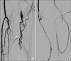
Fig. 1
(A) Fistulographies show an iatrogenic fistula in the needling site of arterial limb, which communicated between the graft and native vein and drained into upper arm vein via multiple collateral veins. (B) After further advancing the catheter, diffuse stenotic venous limb and stenosis of venous anastomosis can be seen.

References
1. You YK, Koh YB. Clinical experience of PTEF (polytetrafluoroethylene) graft A-V fistula for hemodialysis of chronic renal failure patient. J Korean Surg Soc. 1991. 41:345–351.
2. Wilson SE. Wilson SE, editor. Complications of vascular access procedures. Vascular Access: Principles and Practice. 1996. 3rd ed. St. Louis: Mosby;189–203.
3. . III. NKF-K/DOQI Clinical Practice Guidelines for Vascular Access: update 2000. Am J Kidney Dis. 2001. 37(1):supple 1. S137–S181.
4. Min SK, Park YH, Lee HH, Lee JS, Chung WK, Lee JH, et al. Iatrogenic fistula between prosthetic haemodialysis access graft and autogenous vein: unusual cause of graft thrombosis. Nephrol Dial Transplant. 2004. 19:2647–2649.
5. Davidson IJA. Access for Dialysis: Surgical and Radiologic Procedures. 2002. 2nd ed. Georgetown: Landes Bioscience:
 XML Download