

 PDF
PDF ePub
ePub Citation
 Print
Print


Abstract
Purpose:
To determine the myopic shift and cataract change after lens sparing vitrectomy (LSV) in patients with idiopathic epi-retinal membrane (ERM) in their 5th and 6th decade of life.
Methods:
The medical records of patients undergoing LSV for idiopathic ERM from 2008 to 2012 were reviewed. Patients with previous intraocular surgery, preoperative significant cataract, under 40 or over 60 years of age and a follow-up period of less than 6 months were excluded. The change in refractive errors, visual acuity, and cataract grade were evaluated for 6 months af-ter LSV as well as the correlation between myopic shift and cataract change at 6 months after LSV.
Results:
Twenty-eight eyes were included in this study. The cataract status worsened by 1.07 levels, myopia progressed by 3.13 diopters ( p < 0.001), and uncorrected visual acuity (log MAR) decreased from 0.73 to 0.98 ( p = 0.022) at 6 months after LSV. Additional cataract surgery was performed in 22 eyes (78.6%) at 13.6 months on average during the follow-up period (average 16.9 months). In 6 eyes (21.4%) having myopic change below 1.0 diopters, crystalline lens was preserved for 36 months after vitrectomy. Conversely, 22 eyes (78.6%) with myopic change over 1.5 diopters required cataract surgery. Therefore, myopic change over 1.5 diopters could be a major postoperative change predicting the necessity for cataract surgery ( p < 0.001).
Conclusions:
LSV for ERM caused a significant myopic shift and cataract changes in patients in their 5th and 6th decade of life and additional cataract surgery was required in 78.6% of patients within 3 years after vitrectomy. The myopic shift over 1.5 diop-ters at 6 months after vitrectomy could be a major postoperative change predicting the necessity for cataract surgery.
References
1. Mitchell P, Smith W, Chey T, et al. Prevalence and associations of epiretinal membranes. The Blue Mountains Eye Study, Australia. Ophthalmology. 1997; 104:1033–40.
2. Fraser-Bell S, Guzowski M, Rochtchina E, et al. Five-year cumu-lative incidence and progression of epiretinal membranes: the Blue Mountains Eye Study. Ophthalmology. 2003; 110:34–40.
3. Klein R, Klein BE, Wang Q, Moss SE. The epidemiology of epi-retinal membranes. Trans Am Ophthalmol Soc. 1994; 92:425–30.
4. de Bustros S, Thompson JT, Michels RG, et al. Vitrectomy for idio-pathic epiretinal membranes causing macular pucker. Br J Ophthalmol. 1988; 72:692–5.

5. de Bustros S, Rice TA, Michels RG, et al. Vitrectomy for macular pucker: use after treatment of retinal tears or retinal detachment. Arch Ophthalmol. 1988; 106:758–60.
6. Grewing R, Mester U. Results of surgery for epiretinal membranes and their recurrences. Br J Ophthalmol. 1996; 80:323–6.
7. Hsuan JD, Brown NA, Bron AJ, et al. Posterior subcapsular and nuclear cataract after vitrectomy. J Cataract Refract Surg. 2001; 27:437–44.
8. Melberg NS, Thomas MA. Nuclear sclerotic cataract after vi-trectomy in patients younger than 50 year of age. Ophthalmology. 1995; 102:1466–71.
9. Thompson JT. The role of patient age and intraocular gas use in cat-aract progression after vitrectomy for macular holes and epiretinal membranes. Am J Ophthalmol. 2004; 137:250–7.
10. Dugas B, Ouled-Moussa R, Lafontaine PO, et al. Idiopathic epi-retinal macular membrane and cataract extraction: combined ver-sus consecutive surgery. Am J Ophthalmol. 2010; 149:302–6.
11. Treumer F, Bunse A, Rudolf M, Roider J. Pars plana vitrectomy, phacoemulsification and intraocular lens implantation. Compar-ison of clinical complications in a combined versus two-step surgi-cal approach. Graefes Arch Clin Exp Ophthalmol. 2005; 244:808–15.
12. Jung KI, Song MH, Roh YJ. Combined clear corneal phacoemulsi-fication and vitrectomy versus two-step surgery in Korean patients with idiopathic epiretinal membrane. J Korean Ophthalmol Soc. 2011; 52:203–9.
13. Lahey JM, Francis RR, Kearney JJ. Combining phacoemulsifica-tion with pars plana vitrectomy in patients with proliferative dia-betic retinopathy: a series of 223 cases. Ophthalmology. 2003; 110:1335–9.
14. Chylack LT Jr, Wolfe JK, Singer DM. . The Lens Opacities Classification System III. The Longitudinal Study of Cataract Study Group. Arch Ophthalmol. 1993; 111:831–6.
15. Michels RG, Ryan SJ Jr. Results and complications of 100 consec-utive cases of pars plana vitrectomy. Am J Ophthalmol. 1975; 80:24–9.
16. Shui YB, Holekamp NM, Kramer BC, et al. The gel state of the vit-reous and ascorbate-dependent oxygen consumption: relationship to the etiology of nuclear cataracts. Arch Ophthalmol. 2009; 127:475–82.
17. Holekamp NM, Bai F, Shui YB, et al. Ischemic diabetic retinop-athy may protect against nuclear sclerotic cataract. Am J Ophthalmol. 2010; 150:543–50.e1.
18. Sawa M, Ohji M, Kusaka S, et al. Nonvitrectomizing vitreous sur-gery for epiretinal membrane: long-term follow-up. Ophthalmol-ogy. 2005; 112:1402–8.
19. Fujikado T, Kuroda T, Ninomiya S, et al. Age-related changes in ocular and corneal aberrations. Am J Ophthalmol. 2004; 138:143–6.
20. Kawakubo H, Sato Y, Shimada H, et al. Myopic change in re-fraction due to nuclear sclerotic changes after vitreous surgery. Folia Ophthalmol Jpn. 1996; 47:396–400.
21. Brown NA, Hill AR. Cataract: the relation between myopia and cataract morphology. Br J Ophthalmol. 1987; 71:405–14.
22. Reeves BC, Hill AR, Brown NA. Myopia and cataract. Lancet. 1987; 2:964.
23. Kuroda T, Fujikado T, Maeda N, et al. Wavefront analysis of high-er-order aberrations in patients with cataract. J Cataract Refract Surg. 2002; 28:438–44.
24. Kuroda T, Fujikado T, Maeda N, et al. Wavefront analysis in eyes with nuclear or cortical cataract. Am J Ophthalmol. 2002; 134:1–9.
25. Okamoto Y, Okamoto F, Hiraoka T, Oshika T. Refractive changes after lens-sparing vitrectomy for rhegmatogenous retinal detach-ment. Am J Ophthalmol. 2014; 158:544–9.
26. de Bustros S, Thompson JT, Michels RG, et al. Nuclear sclerosis after vitrectomy for idiopathic epiretinal membranes. Am J Ophthalmol. 1988; 105:160–4.
27. Cherfan GM, Michels RG, de Bustros S, et al. Nuclear sclerotic cataract after vitrectomy for idiopathic epiretinal membranes caus-ing macular pucker. Am J Ophthalmol. 1991; 111:434–8.
28. Saito Y, Lewis JM, Park I, et al. Nonvitrectomizing vitreous sur-gery: a strategy to prevent postoperative nuclear sclerosis. Ophthalmology. 1999; 106:1541–5.
29. Sawa M, Saito Y, Hayashi A, et al. Assessment of nuclear sclerosis after nonvitrectomizing vitreous surgery. Am J Ophthalmol. 2001; 132:356–62.
Figure 1.
Kaplan-Meier survival curve of crystal lens after lens sparing vitrectomy. 78.6% of patients needed additional cataract surgery during 3 years after vitrectomy.
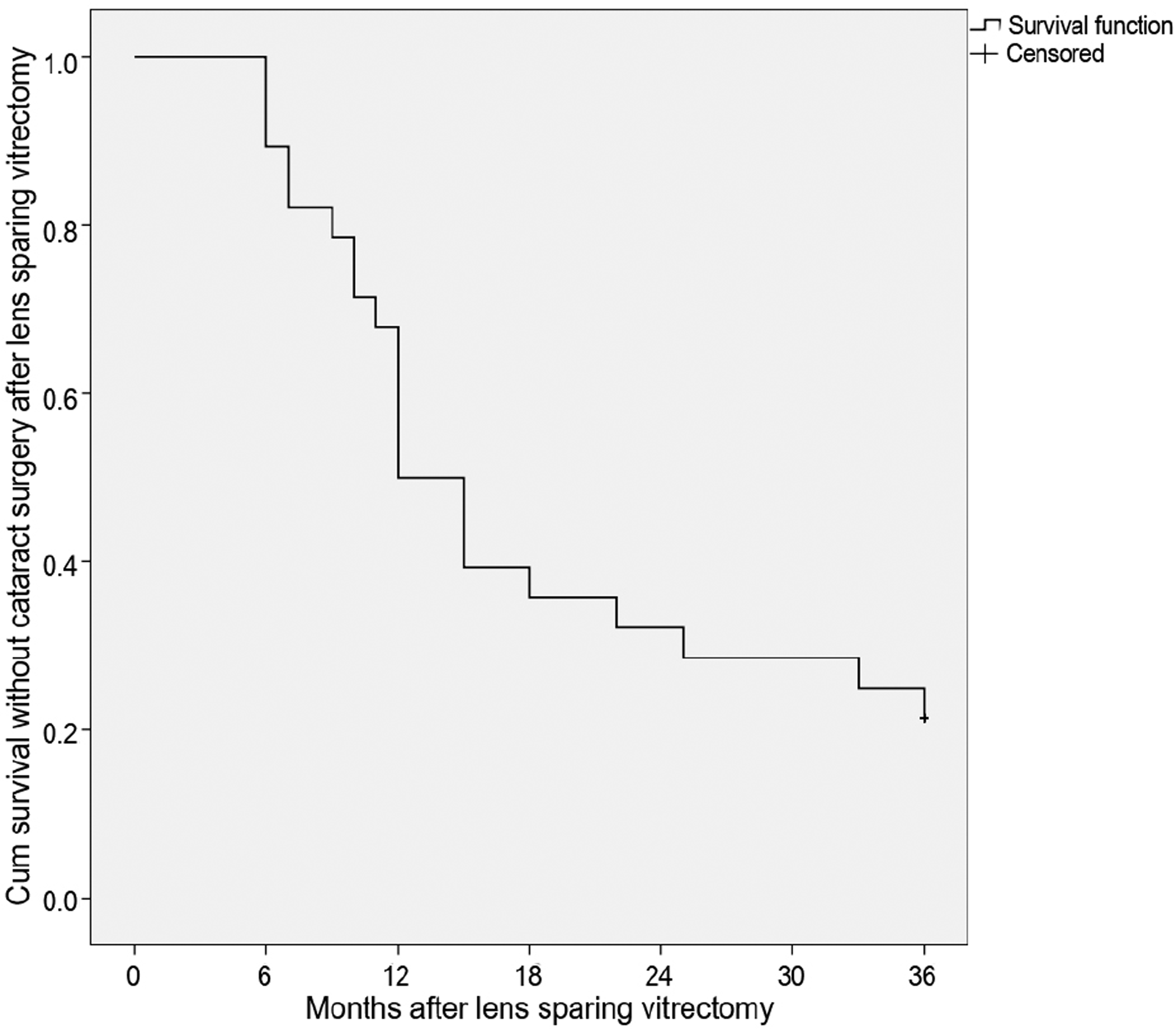
Figure 2.
Kaplan-Meier survival curve of crystal lens accord-ing to age (5th and 6th decade) after lens sparing vitrectomy. There was no significant difference of survival rate between two age groups in 3 years after vitrectomy ( p = 0.288 by Fisher exact test).
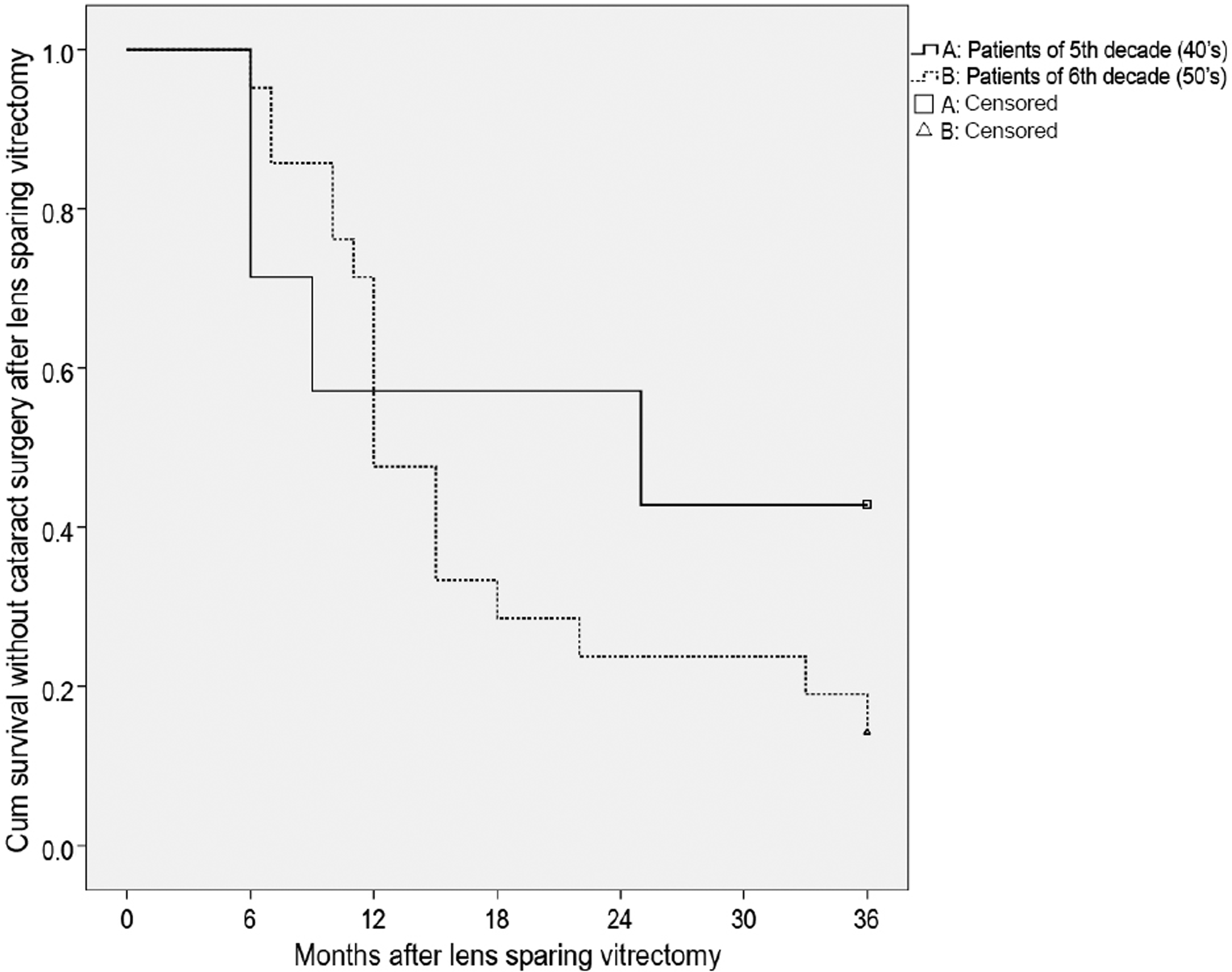
Table 1.
Comparisons of data between baseline and 6 months after vitrectomy
| Preoperative value | Postoperative 6-months | p-value | |
|---|---|---|---|
| LOCS III grading of nucleus | 0.9 ± 0.9 | 1.9 ± 1.2 | <0.001* |
| Corrected V.A. (log MAR) | 0.51 ± 0.30 | 0.52 ± 0.35 | 0.471 |
| Uncorrected V.A. (log MAR) | 0.73 ± 0.42 | 0.98 ± 0.50 | 0.022* |
| Spherical equivalent (D) | -0.89 ± 2.76 | -4.02 ± 3.47 | <0.001* |
| CSMT (um) | 499.0 ± 99.6 | 360.6 ± 41.7 | <0.001∗ |
Table 2.
Differences according to myopic shift at 6 months after vitrectomy
| Myopic shift less than -1.0 Diopters | Myopic shift more than -1.5 Diop | ters p-value | |
|---|---|---|---|
| Number of patients | 6 (21.4) | 22 (78.6) | |
| Age (years) | 53.0 ± 4.4 | 52.0 ± 2.0 | 0.259 |
| Corrected V.A. (log MAR) | 0.28 ± 0.16 | 0.5 ± 0.37 | 0.065 |
| Uncorrected V.A. (log MAR) | 0.52 ± 0.32 | 0.95 ± 0.46 | 0.007* |
| Spherical equivalent (Diopter) | -0.74 ± 1.12 | -3.69 ± 3.35 | <0.001* |
| CSMT (um) | 372.5 ± 39.1 | 351.7 ± 42.6 | 0.309 |
| Worsened cataract grade | 2 (33.3) | 20 (90.9) | 0.010* |
| Cataract operation during 3 years | 0 (0) | 22 (100) | <0.001∗ |
 XML Download