

 PDF
PDF ePub
ePub Citation
 Print
Print


Abstract
Case summary
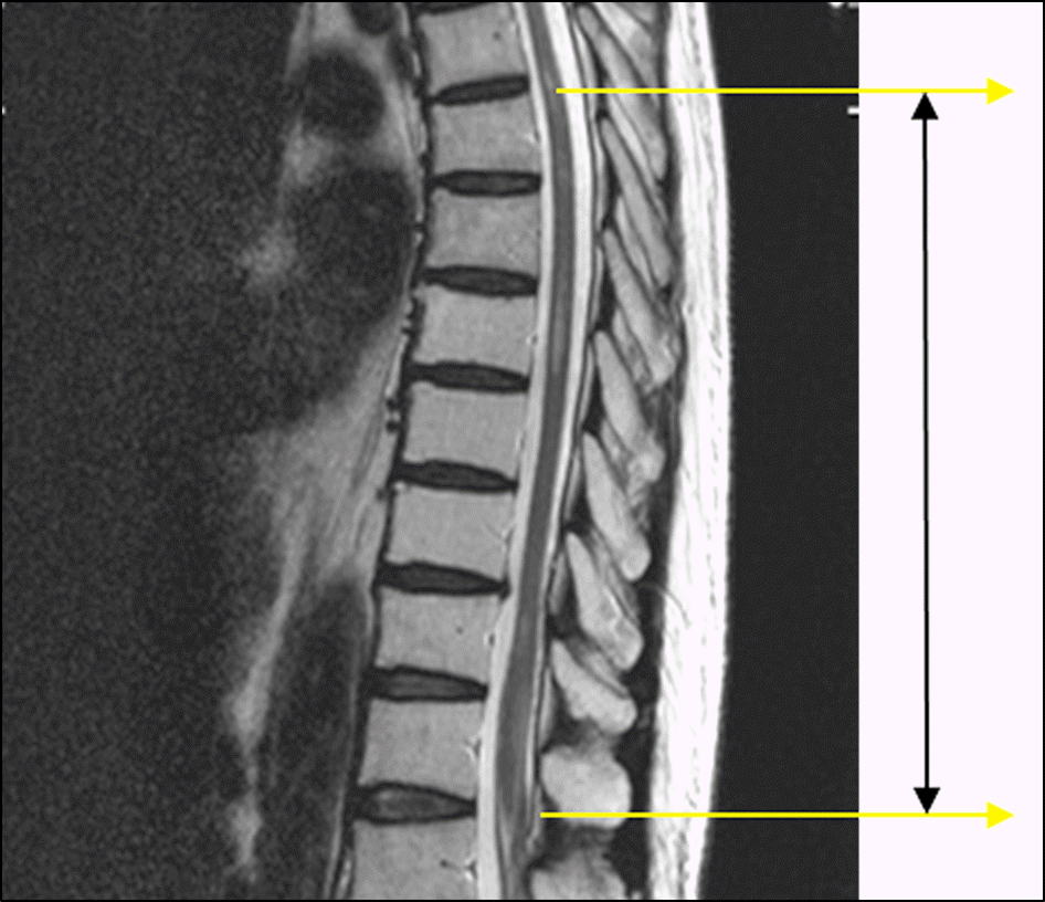
A 70-year-old male with hepatocellular carcinoma presented with bilateral visual loss. A relative afferent pupillary defect was not observed in either eye due to bilateral mydriasis. On brain MRI, there was no specific finding, however, on spine MRI, multiple and severe myelopathies were observed. After high-dose methylprednisolone pulse therapy, the visual acuity was 0.03 in the right eye and counting fingers at 30 cm in the left eye.
References
1. Wingerchuck DM, Hogancamp WF, O'Brien PC, Weinshenker BG. The clinical course of neuromyelitis optica(Devic's syndrome). Neurology. 1999; 53:1107–14.
2. Lennon VA, Wingerchuck DM, Kryzer TJ. . A serum autoantibody marker of neuromyelitis optica: distinction from multiple sclerosis. Lancet. 2004; 364:2106–12.

3. Pau D, Zubidi NA, Yalamanchili S. . Optic neuritis. Eye (Lond). 2011; 25:833–42.
4. Wingerchuck DM, Lennon VA, Pittock SJ. . Revised diagnostic criteria for neuromyelitis optica. Neurology. 2006; 66:1485–9.
5. Wingerchuck DM, Lennon VA, Lucchinetti CF. . The spectrum of neuromyelitis optica. Lancet Neurol. 2007; 6:805–15.
6. Noh Y, Kang EH, Hwang JM, Kim JS. Bilateral optic neuritis as the first manifestation of systemic lupus erythematous associated with neuromyelitis optica. J Korean Neurol Assoc. 2010; 28:323–5.
7. Pittock SJ, Lennon VA. Aquaporin-4 autoantibodies in a paraneoplastic context. Arch Neurol. 2008; 65:629–32.
8. Darnell RB, Posner JB. Paraneoplastic syndromes involving the nervous system. N Engl J Med. 2003; 349:1543–54.
9. Kitazawa Y, Warabi Y, Bandoh M. . Eldery-onset neuromyelitis optica which developed after diagnosis of prostate adenocarcinoma. Intern Med. 2012; 51:103–7.
Figure 1.
Liver CT scan of the patient at the initial visit. About 5.3 × 5.0 cm-sized round mass in the left lobe of the liver (expanding nodular type).
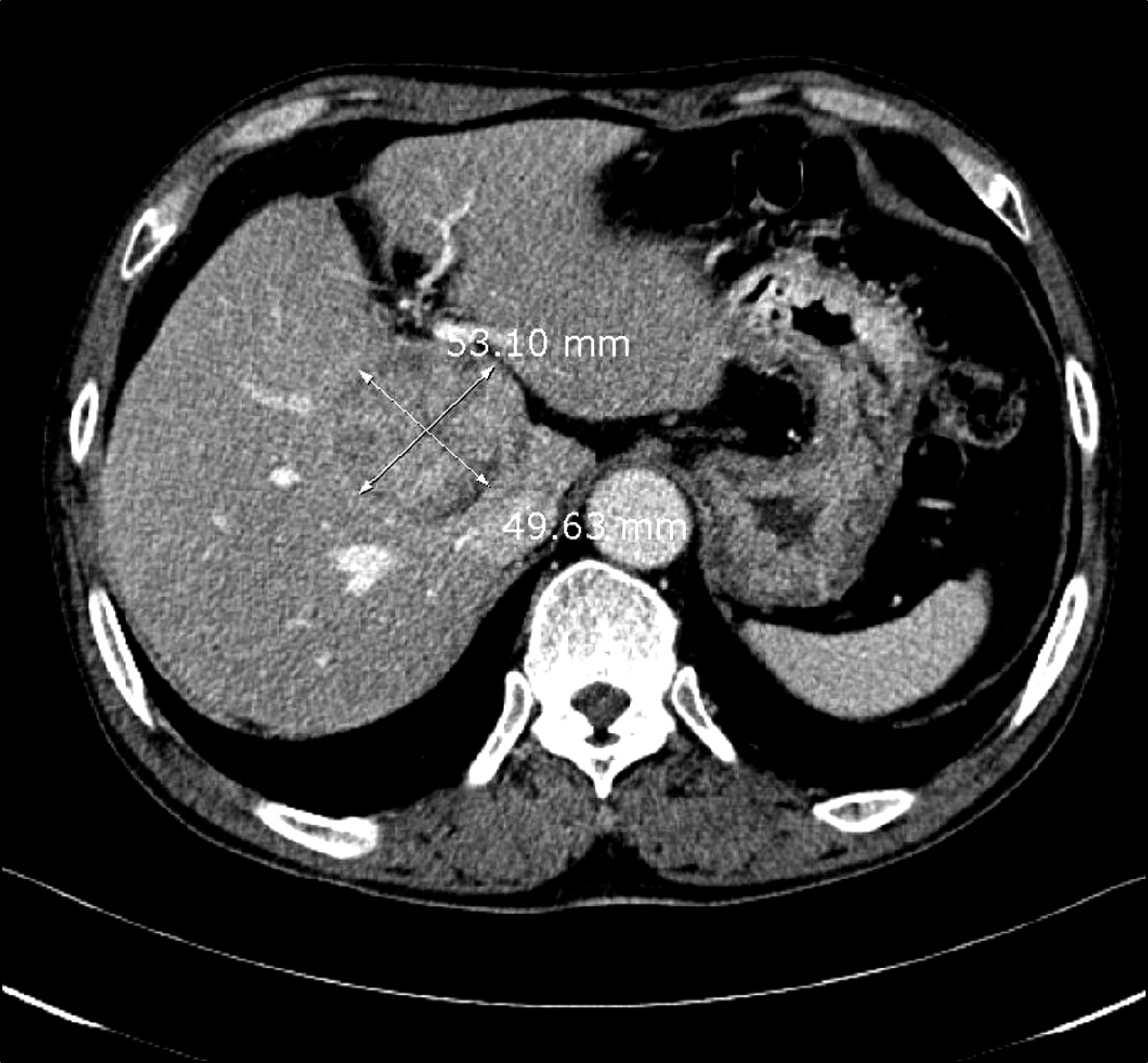
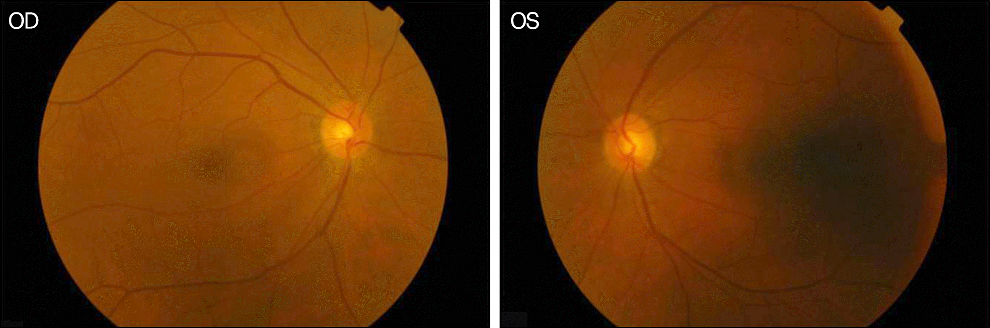
Figure 3.
Fundus photographs at the initial presentation. There are no specific abnormalities in the retina of both eyes.
 XML Download