

 PDF
PDF ePub
ePub Citation
 Print
Print


Abstract
Purpose
To measure choroidal thickness in healthy myopic eyes and to evaluate the relationship among choroidal thickness and refractive power and axial length.
Methods
Eighty healthy myopic eyes were evaluated in the present study. The refractive power was measured using an automatic refractor and the axial length using A-scan. The subjects were divided into two groups based on refractive power (≥-6.0 D and <-6.0 D) and axial length (≥25 mm and <25 mm). The choroidal thickness was measured using spectral domain (SD) optical coherence tomography (3-dimensional [3D] OCT-2000, Software Version 6.01; Topcon Corp., Tokyo, Japan), and the statistical relationship between the two groups was analyzed.
Figures and Tables
Figure 1
Scatter plots and regression line of the choroidal thickness against refractive power. p < 0.001; y = 14.19 ± 382.11; R2 = 0.266.
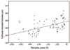
Figure 2
Scatter plots and regression line of the choroidal thickness against axial length. p < 0.001; y = -27.87 ± 1025.61; R2 = 0.197.
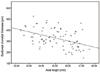
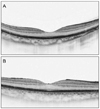
Figure 3
Comparison between OCT scanning in two eyes of different refractive powers and axial length. A 32-year-old man (refractive power -8.25 D, axial length 27.57 mm) with a subfoveal choroidal thickness of 227 µm (A) and a 33-year-old man (refractive power -2.875D, axial length 24.89 mm) with subfoveal choroidal thickness of 333 µm (B).


References
1. Puliafito CA, Hee MR, Lin CP, et al. Imaging of macular diseases with optical coherence tomography. Ophthalmology. 1995. 102:217–229.
2. Muscat S, Parks S, Kemp E, Keating D. Repeatability and reproducibility of macular thickness measurements with the Humphrey OCT system. Invest Ophthalmol Vis Sci. 2002. 43:490–495.
3. Margolis R, Spaide RF. A pilot study of enhanced depth imaging optical coherence tomography of the choroid in normal eyes. Am J Ophthalmol. 2009. 147:811–815.
4. Ikuno Y, Kawaguchi K, Nouchi T, Yasuno Y. Choroidal thickness in healthy Japanese subjects. Invest Ophthalmol Vis Sci. 2010. 51:2173–2176.
5. Manjunath V, Taha M, Fujimoto JG, Duker JS. Choroidal thickness in normal eyes measured using Cirrus HD optical coherence tomography. Am J Ophthalmol. 2010. 150:325–329.
6. Imamura Y, Fujiwara T, Margolis R, Spaide RF. Enhanced depth imaging optical coherence tomography of the choroid in central serous chorioretinopathy. Retina. 2009. 29:1469–1473.
7. Maruko I, Iida T, Sugano Y, et al. Subfoveal choroidal thickness after treatment of Vogt-Koyanagi-Harada disease. Retina. 2011. 31:510–517.
8. Ikuno Y, Tano Y. Retinal and choroidal biometry in highly myopic eyes with spectral-domain optical coherence tomography. Invest Ophthalmol Vis Sci. 2009. 50:3876–3880.
9. Fujiwara T, Imamura Y, Margolis R, et al. Enhanced depth imaging optical coherence tomography of the choroid in highly myopic eyes. Am J Ophthalmol. 2009. 148:445–450.
10. Avila MP, Weiter JJ, Jalkh AE, et al. Natural history of choroidal neovascularization in degenerative myopia. Ophthalmology. 1984. 91:1573–1581.
11. Curtin BJ. Myopia: a review of its etiology, pathogenesis, and treatment. Surv Ophthalmol. 1970. 15:1–17.
12. Curtin BJ. Posterior staphyloma development in pathologic myopia. Ann Ophthalmol. 1982. 14:655–658.
13. Grossniklaus HE, Green WR. Pathologic findings in pathologic myopia. Retina. 1992. 12:127–133.
14. Kim SW, Oh J, Kwon SS, et al. Comparison of choroidal thickness among patients with healthy eyes, early age-related maculopathy, neovascular age-related macular degeneration, central serous chorioretinopathy, and polypoidal choroidal vasculopathy. Retina. 2011. 31:1904–1911.
15. Jaffe GJ, Caprioli J. Optical coherence tomography to detect and manage retinal disease and glaucoma. Am J Ophthalmol. 2004. 137:156–169.
16. Spaide RF, Koizumi H, Pozzoni MC. Enhanced depth imaging spectral-domain optical coherence tomography. Am J Ophthalmol. 2008. 146:496–500.
17. Agawa T, Miura M, Ikuno Y, et al. Choroidal thickness measurement in healthy Japanese subjects by three-dimensional high-penetration optical coherence tomography. Graefes Arch Clin Exp Ophthalmol. 2011. 249:1485–1492.
18. Alm A, Bill A. Ocular and optic nerve blood flow at normal and increased intraocular pressures in monkeys (Macaca irus): a study with radioactively labelled microspheres including flow determinations in brain and some other tissues. Exp Eye Res. 1973. 15:15–29.
19. Alm A, Bill A. Moses RA, Hart WM, editors. Ocular circulation. Adlers' Physiology of the Eye. 1987. St. Louis: Mosby.
20. Torczynski E. Duane TD, Jaeger EA, editors. Choroid and suprachoroid. Biomedical Foundations of Ophthalmology. 1987. v. 1. Philadelphia: Harper & Row.
21. Parver LM, Auker C, Carpenter DO. Choroidal blood flow as a heat dissipating mechanism in the macula. Am J Ophthalmol. 1980. 89:641–646.
22. Rohen JW. Bargmann Handbuch der mikroskoplschen Anatomie des Menschen. Haut and Sinnesorgane. IV. Das Auge und seine Hilfsogane. 1964. Berlin: Springer-Verlag.
23. Siam A. Macular hole with central retinal detachment in high myopia with posterior staphyloma. Br J Ophthalmol. 1969. 53:62–63.
24. Curtin BJ, Karlin DB. Axial length measurements and fundus changes of the myopic eye. Am J Ophthalmol. 1971. 71(1 Pt 1):42–53.
25. Esmaeelpour M, Povazay B, Hermann B, et al. Three-dimensional 1060-nm OCT: choroidal thickness maps in normal subjects and improved posterior segment visualization in cataract patients. Invest Ophthalmol Vis Sci. 2010. 51:5260–5266.
26. Agawa T, Miura M, Ikuno Y, et al. Choroidal thickness measurement in healthy Japanese subjects by three-dimensional high-penetration optical coherence tomography. Graefes Arch Clin Exp Ophthalmol. 2011. 249:1485–1492.
 XML Download