

 PDF
PDF ePub
ePub Citation
 Print
Print


Abstract
Purpose
The butterfly-shaped pigment dystrophy is an extremely rare autosomal dominant retinal disorder. The authors present a case of butterfly-shaped pigment dsytrophy not reported previously in Korea.
Case summary
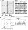
A 45-year-old man had bilateral blurred vision for 2 years. His visual acuity was 0.2 in the right eye, and 0.3 in the left and was uncorrected. Funduscopically, yellow pigment deposits were present at the level of retinal pigment epithelium (RPE) in the central macula of both eyes. Fluorescein angiography (FA) revealed a central, dark, butterfly-shaped lesion surrounded by a region of hyperfluorescence, Spectral domain optical coherence tomography (OCT) showed thick elevation of the RPE with hyperreflectivity and disruption of the inner and outer segment (IS/OS) interface of the photoreceptors. The patient had normal color vision, visual field and electroretinograms and reduced electrooculograms.
Conclusions
In general, butterfly-shaped pigment dystrophy is known to have good visual prognosis. However, in some cases the disease can be a chronic progressive disorder with secondary involvement of the photoreceptors, as exemplified this patient. The authors anticipate more detail regarding the natural course of this disease will be obtained through spectral domain OCT.
Figures and Tables
Figure 1
Fundus photographs (A, B) and fluorescein angiography (C, D) of a case of butterfly-shaped pigment dystrophy. (A, B) He had macular epiretinal membrane and yellow pigment deposits at the level of RPE in the central macula of both eyes. (C, D) The both eyes show some hypofluorescent, radially orientated, macular lesions intermingled with hyperfluorescent areas. Hyofluorescnt areas of fluorescein angiography corresponded to yellowish deposits of the fundus photograph.
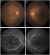
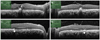
Figure 2
Spectral-domain optical coherence tomography (OCT) findings of the right eye (A, B) and the left eye(C, D) in the patient. (A, C) Horizontal central macular OCTs illustrate thick elevation of retinal pigment epithelium and hyperreflective material deposits in the inner retina. They also show disruption of the inner segment and outer segment (IS/OS) interface at the fovea. (B, D) Horizontal OCTs illustrate elevation of the retinal pigment epithelium. This high reflective signals (arrow) correspond to the accumulation of subretinal yellow material. All the horizontal OCTs show macular epiretinal membrane.
References
1. Deutman AF, van Blommestein JD, Henkes HE, et al. Butterfly-shaped pigment dystrophy of the fovea. Arch Ophthalmol. 1970. 83:558–569.
2. Tuppurainen K, Mäntyjärvi M. The importance of fluorescein angiography in diagnosing pattern dystrophies of the retinal pigment epithelium. Doc Ophthalmol. 1994. 87:233–243.
3. Zhang K, Garibaldi DC, Li Y, et al. Butterfly-shaped pattern dystrophy: a genetic, clinical, and histopathological report. Arch Ophthalmol. 2002. 120:485–490.
4. Zhang K, Nguyen TH, Crandall A, Donoso LA. Genetic and molecular studies of macular dystrophies: recent developments. Surv Ophthalmol. 1995. 40:51–61.
5. van Lith-Verhoeven JJ, Cremers FP, van den Helm B, et al. Genetic heterogeneity of butterfly-shaped pigment dystrophy of the fovea. Mol Vis. 2003. 9:138–143.
6. Isaac DL, Santos RA, Avila M. [Butterfly-shaped pattern dystrophy: case report]. Arq Bras Oftalmol. 2007. 70:129–132.
 XML Download