

 PDF
PDF ePub
ePub Citation
 Print
Print


Abstract
Purpose
To report two cases of trabeculectomy with biodegradable collagen material conducted on two post-vitrectomy patients.
Case summary
The first patient was a 43-year-old woman with uncontrolled increased intraocular pressure (IOP) after pars plana vitrectomy with scleral buckling for diabetic retinopathy and vitreous hemorrhage. Another patient, a 28-year-old woman with aphakia, also had uncontrolled increased IOP after pars plana vitrectomy with encircling scleral buckling for retinal detachment. For both of these patients, we performed trabeculectomy using mitomycin C and recently developed biodegradable collagen matrix. In the first case, the IOP was increased three months after the surgery, for which needling was done. After needling, the IOP was well controlled in the range of 16 to 19 mmHg up to eight months postoperatively with stilling anti-glaucomatous eyedrops (Cosopt®, Xalatan®). In the second case, IOPwas increased one week after the surgery, but it was well controlled between 14 to 21 mmHg up to nine months postoperatively with anti-glaucomatous eye drops (Combigan®).
References
1. Teng CC, Chi HH, Katzin HM. Histology and mechanism of filtering operations. Am J Ophthalmol. 1959; 47:16–33.

2. Addicks EM, Quigley HA, Green WR, Robin AL. Histologic characteristics of filtering blebs in glaucomatous eyes. Arch Ophthalmol. 1983; 101:795–8.
3. Kitazawa Y, Kawase K, Matsuhita H, Minobe M. Trabeculectomy with Mitomycin. Arch Ophthalmol. 1991; 109:1693–8.
4. Skuta GL, Beeson CC, Hlgginbotham EJ, et al. Intraoperative mitomycin versus postoperative 5-fluorouracil in high risk glaucoma filtering surgery. Ophthalmology. 1992; 99:438–4.
5. Cairns JE. Trabeculectomy-preliminary report of a new method. Am J Ophthalmol. 1968; 66:673–9.
6. Chen HS, Ritch R, Krupin T, Hsu WC. Control of filtering bleb structure through tissue bioengineering: An animal model. Invest Ophthalmol Vis Sci. 2006; 47:5310–4.
7. Dietlein TS, Jordan J, Lueke C, Krieglstein GK. Modern concepts in antiglaucomatous implant surgery. Graefes Arch Clin Exp Ophthalmol. 2008; 246:1653–64.
8. Aptel F, Dumas S, Denis P. Ultrasound biomicroscopy and optical coherence tomography imaging of filtering blebs after deep sclerectomy with new collagen implant. Eur J Ophthalmol. 2009; 19:223–30.

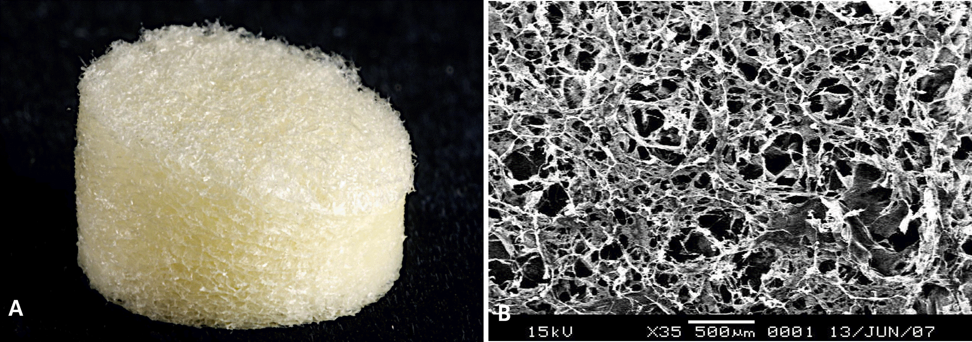
Figure 2.
Trabeculectomy with mitomycin C (MMC) using biodegradable material of Case 1. (A) Scleral flap formation. (B) MMC soaking under scleral flap. (C) Iridectomy. (D) Positioning of biodegradable collagen material. (E) Conjunctival suture. (F) Viscoelastic injection to reform anterior chamber.
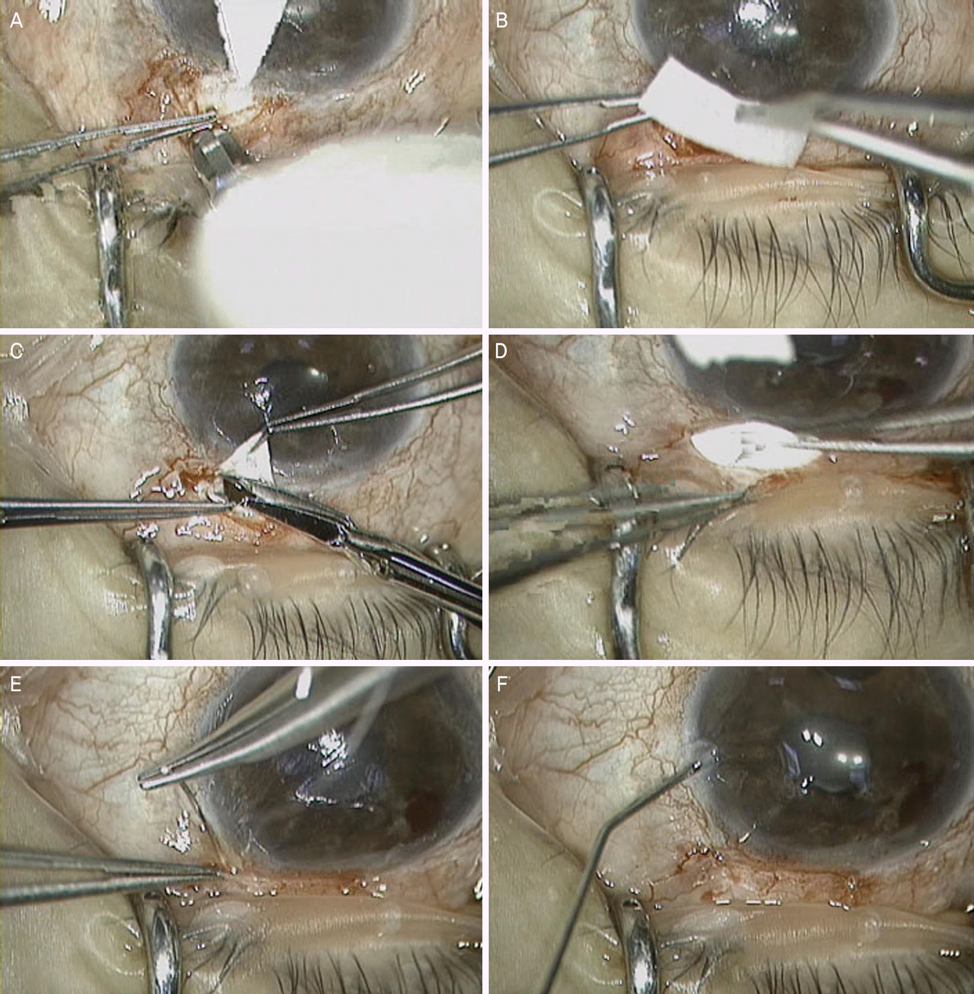
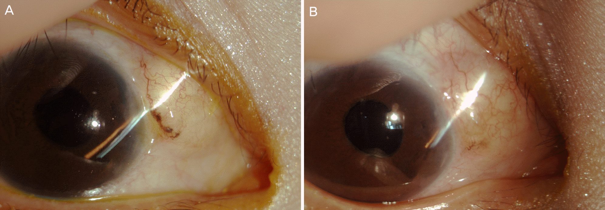
Figure 3.
Slit lamp photograph of Case 1. (A) Highly elevated bleb is observed 1 day postoperatively. (B) Well elevated bleb is observed 8 months postoperatively.
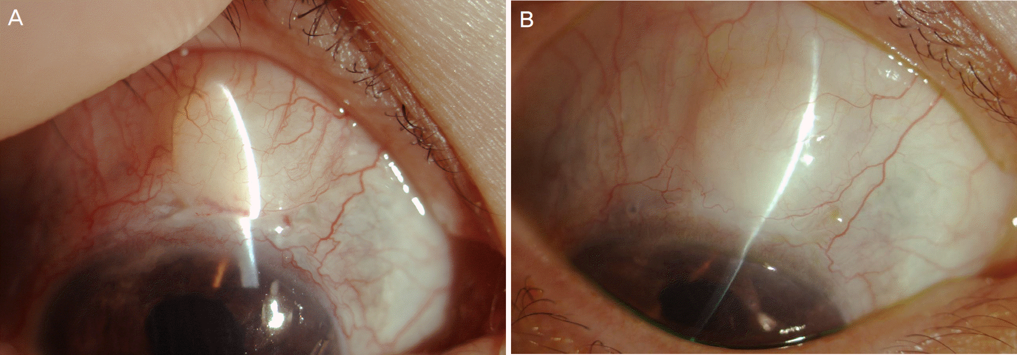
Figure 4.
Each patient’s intraocular pressure changes (Arrow: postoperative 1 day of Case 2, due to hypotony, anterior chamber was reformed with BSS solution and additional scleral suture and collagen implantation also done. Arrow head: postoperative 3 months of Case 1, needling of bleb was done). d=day; w=week; m=month.
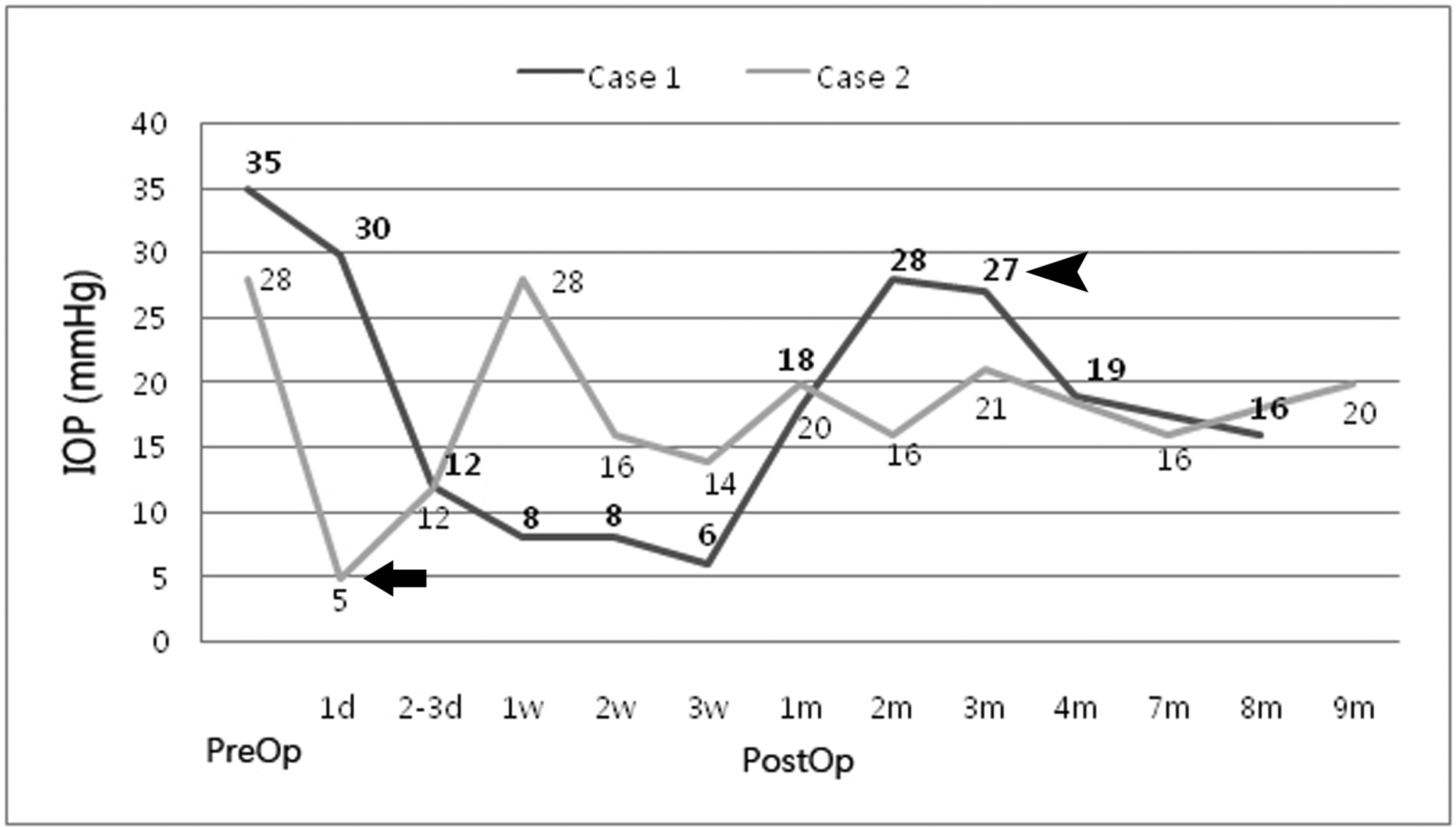
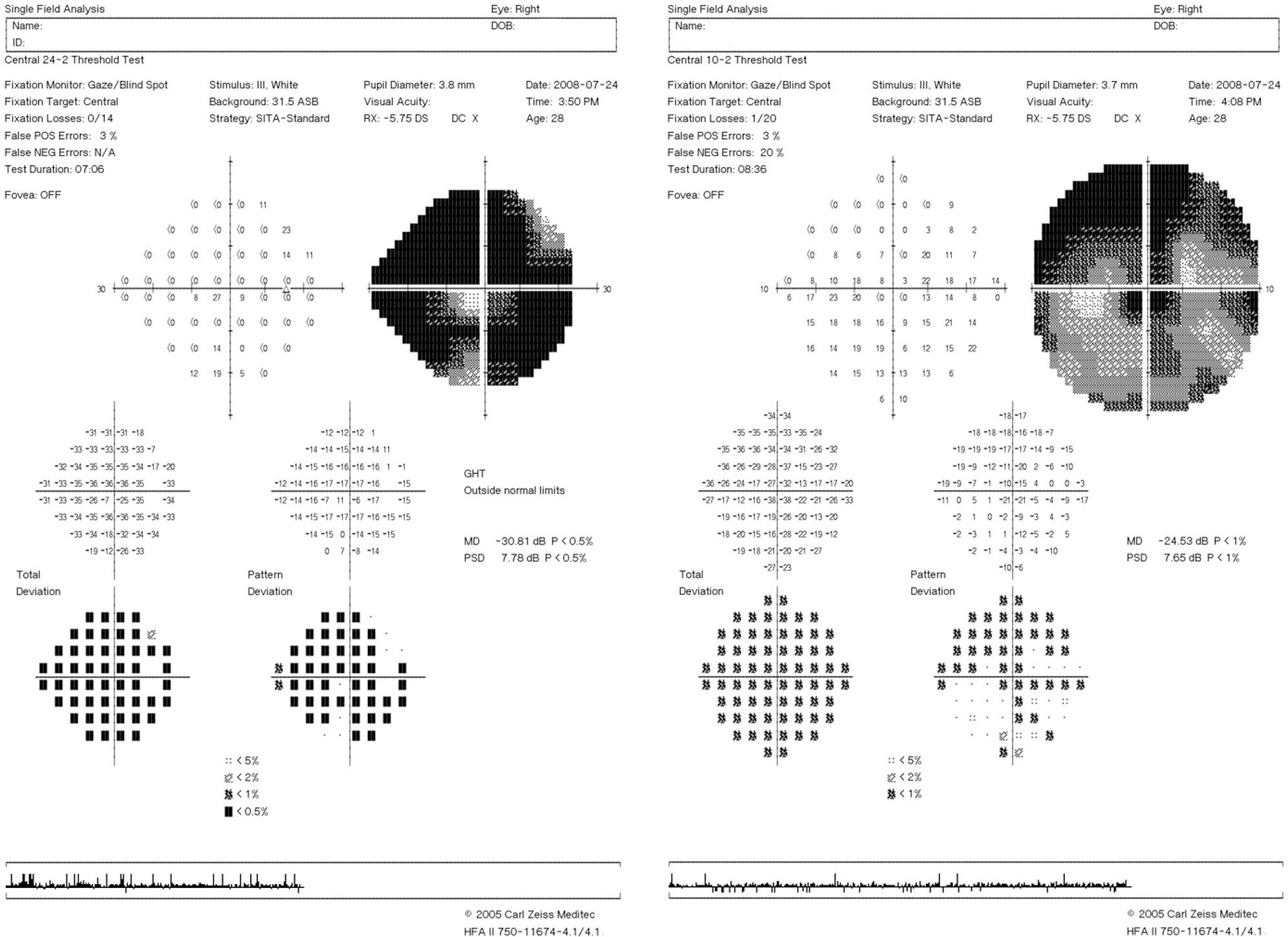
Figure 5.
Initial perimetry (Humphrey 24-2, central 10-2 SITA) of Case 2. It shows only central island is saved.
 XML Download