

 PDF
PDF ePub
ePub Citation
 Print
Print


Abstract
Case summary
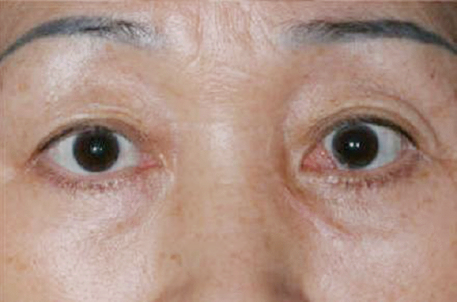
A 60-year-old woman was referred for evaluation of proptosis of the left eye, which had developed about 2 years earlier. Upon initial examination, a movable mass was palpated in the medial aspect of the left orbit. Magnetic resonance imaging of the orbit showed a 2.5 cm-sized, ovoid, cystic mass located between the left eyeball and the medial wall of the orbit. Excisional biopsy of the orbital mass was performed. The orbital mass was a well-circumscribed cystic lesion, adherent to the medial rectus muscle. Histological examination revealed that the cyst was lined with multiple layers of cuboidal epithelium with goblet cells. A diagnosis of primary conjunctival cyst was made.
References
1. Shields JA, Kaden IH, Eagle RC Jr, Shields CL. Orbital dermoid cysts: clinicopathologic correlations, classification, and management. The 1997 Josephine E. Schueler Lecture. Ophthal Plast Reconstr Surg. 1997; 13:265–76.
2. Jakobiec FA, Bonanno PA, Sigelman J. Conjunctival adnexal cysts and dermoids. Arch Ophthalmol. 1978; 96:1404–9.

3. Goldstein MH, Soparkar CN, Kersten RC, et al. Conjunctival cysts of the orbit. Ophthalmology. 1998; 105:2056–60.
4. Shields JA, Shields CL. Orbital cysts of childhood-classification, clinical features, and management. Surv Ophthalmol. 2004; 49:281–99.
5. Rose GE, O’Donnell BA. Congenital orbital cysts associated with the common sheath of superior rectus and levator palpebrae superioris muscles. Ophthalmology. 1995; 102:135–8.
6. McCarthy RW, Beyer CK, Dallow RL, et al. Conjunctival cysts of the orbit following enucleation. Ophthalmology. 1981; 88:30–5.
7. Boynton JR, Searl SS, Ferry AP, et al. Primary nonkeratinized epithelial (‘conjunctival’) orbital cysts. Arch Ophthalmol. 1992; 110:1238–42.
8. Soll SM, Lisman RD, Harrison W, Weiner M. Conjunctival orbital cyst. Ophthal Plast Reconstr Surg. 1994; 10:216–9.
9. Metz HS, Searl S, Rosenberg P, Sterns G. Giant orbital cyst after strabismus surgery. J AAPOS. 1999; 3:185–7.
10. Imaizumi M, Nagata M, Matsumoto CS, et al. Primary conjunctival epithelial cyst of the orbit. Int Ophthalmol. 2007; 27:269–71.
11. Colombo F, Holbach LM, Naumann GO. Conjunctival cyst and conjunctival dermoid of the orbit. Orbit. 2000; 19:13–9.
12. Owji N, Aslani A. Conjunctival cysts of the orbit after enucleation: the use of trichloroacetic acid. Ophthal Plast Reconstr Surg. 2005; 21:264–6.
13. De Potter P, Kunin AW, Shields CL, et al. Massive orbital cyst of the lateral rectus muscle after retinal detachment surgery. Ophthal Plast Reconstr Surg. 1993; 9:292–6.
14. Johnson DW, Bartley GB, Garrity JA, Robertson DM. Massive epi-thelium-lined inclusion cysts after scleral buckling. Am J Ophthalmol. 1992; 113:439–42.
15. Cibis GW, Waeltermann JM. Muscle inclusion cyst as a complication of strabismus surgery. Am J Ophthalmol. 1985; 100:740–1.
16. Harris GJ, Beatty RL, Massaro BM, Lewandowski MF. Conjunctival implantation cyst of the orbit. Transient visual loss with pregnancy. Arch Ophthalmol. 1989; 107:924.
17. Ho VT, Rao VM, Flanders AE. Postsurgical conjunctival epithelial cysts. AJNR Am J Neuroradiol. 1994; 15:1181–3.
18. Kushner BJ. Subconjunctival cysts as a complication of strabismus surgery. Arch Ophthalmol. 1992; 110:1243–5.
19. Hornblass A, Bosniak S. Orbital cysts following enucleation: the use of absolute alcohol. Ophthalmic Surg. 1981; 12:123–6.
Figure 1.
Clinical photograph showing movable mass in the inferonasal part of the left orbit (arrows).
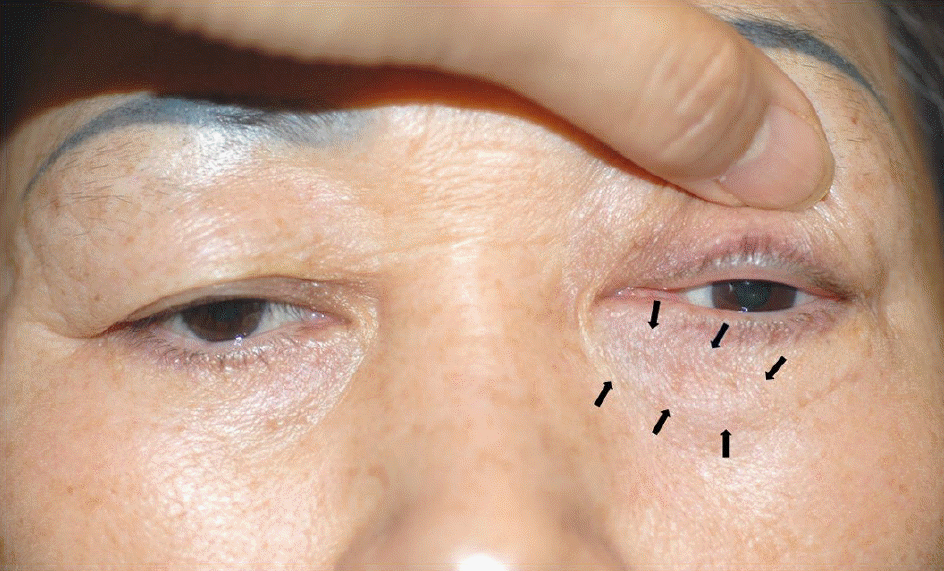
Figure 2.
Axial (A) and coronal (B) CT images reveal a 2.5×2.4×1.4-cm, ovoid, cystic mass in the medial aspect of the left orbit. Lateral displacement of the left globe and pressure remodeling of the medial wall in the left orbit are shown.
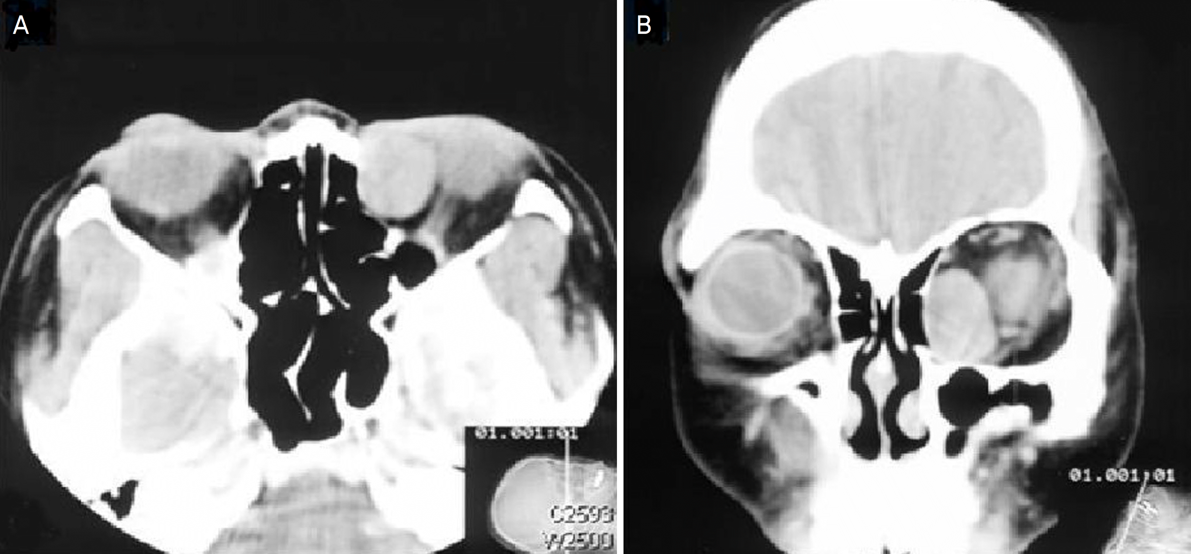
Figure 3.
Axial (A) T1-weighted MR image and coronal (B) T2-weighted MR image demonstrate a well-circum-scribed cystic lesion between the eyeball and medial wall in the left orbit. Axial (C) and sagittal (D) fat-sup-pressed contrast-enhanced T1-weighted MR images show the cystic mass which is adherent to medial rectus muscle without enhancement.
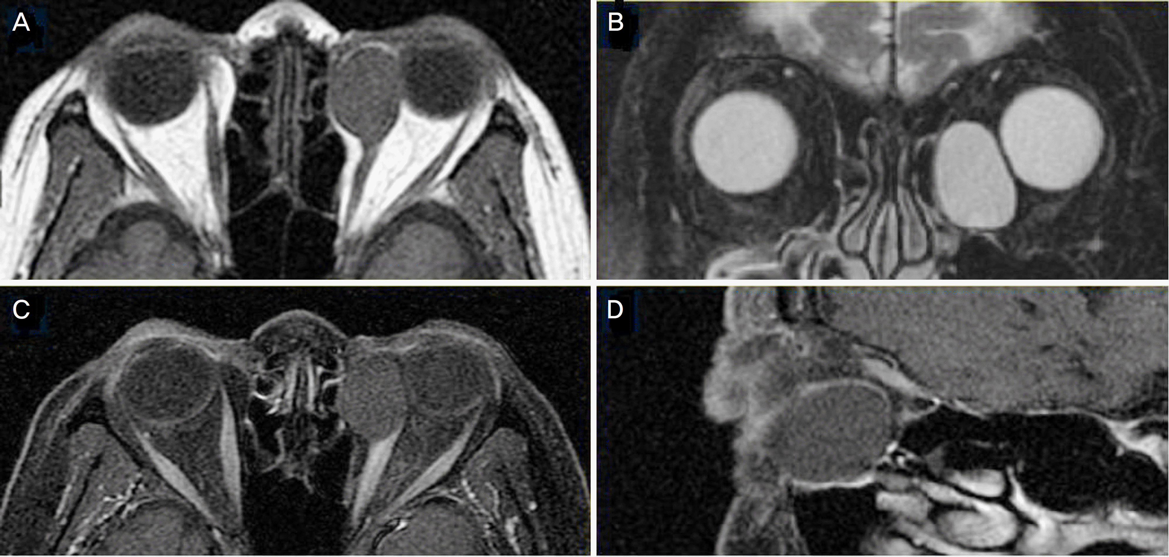
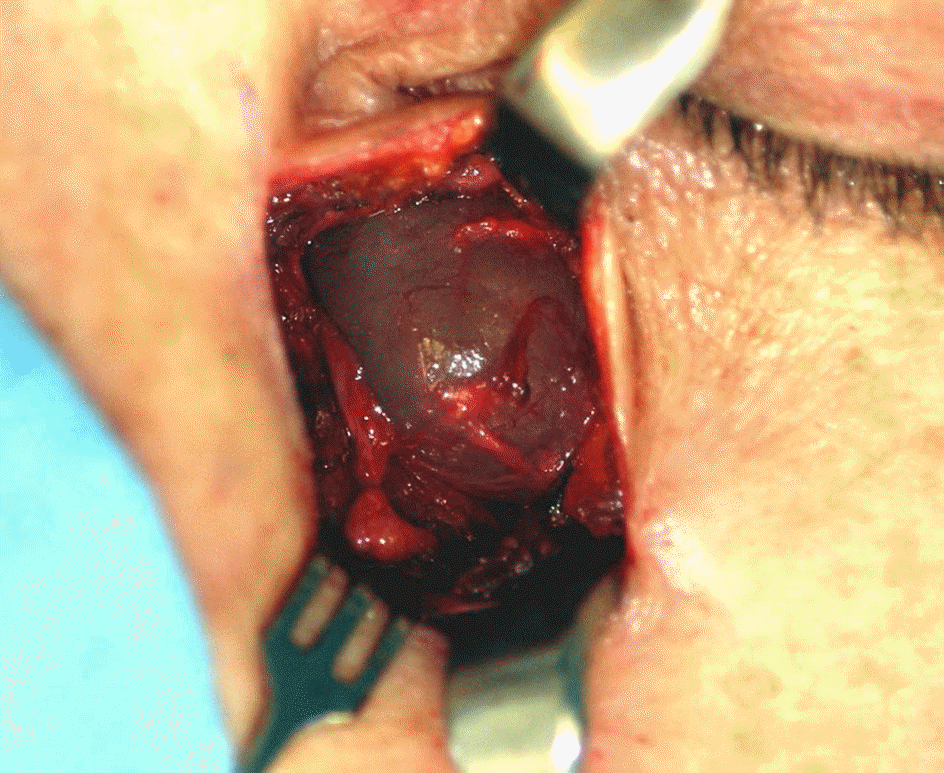
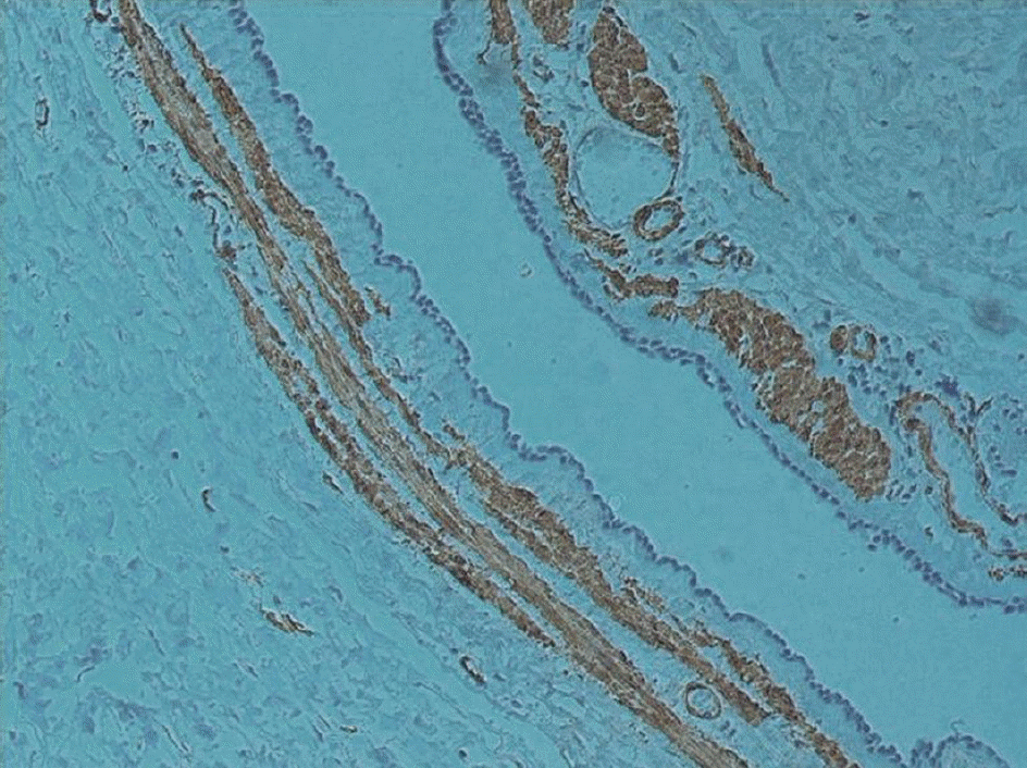
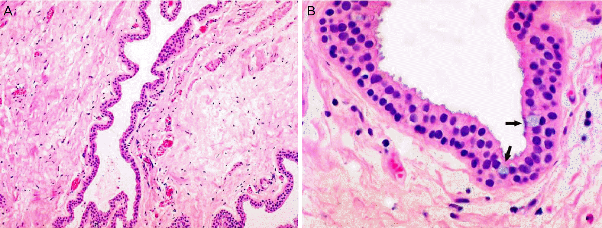
Figure 5.
(A) The cystic wall is lined with nonkeratinized, cuboidal, or columnar epithelium with goblet cells (H & E stain, ×100). (B) Higher magnification showed stratified cuboidal or columnar epithelial cells and goblet cells (arrows) (H & E stain, ×400).
 XML Download