

 PDF
PDF ePub
ePub Citation
 Print
Print


Abstract
Purpose
A Solitary fibrous tumor is a rare orbital neoplasm derived from mesenchymal cells. The neoplasm should be considered in differential diagnosis of any orbital tumor, and immunohistochemial analysis is important for correct diagnosis. The authors herein describe a case of a solitary fibrous tumor in addition to the findings of a literature review. Solitary fibrous tumors can develop from not only the lacrimal gland, but also orbital soft tissue. Until now, there has been no report of a solitary fibrous tumor arising from orbital soft tissue in Korea.
Case summary
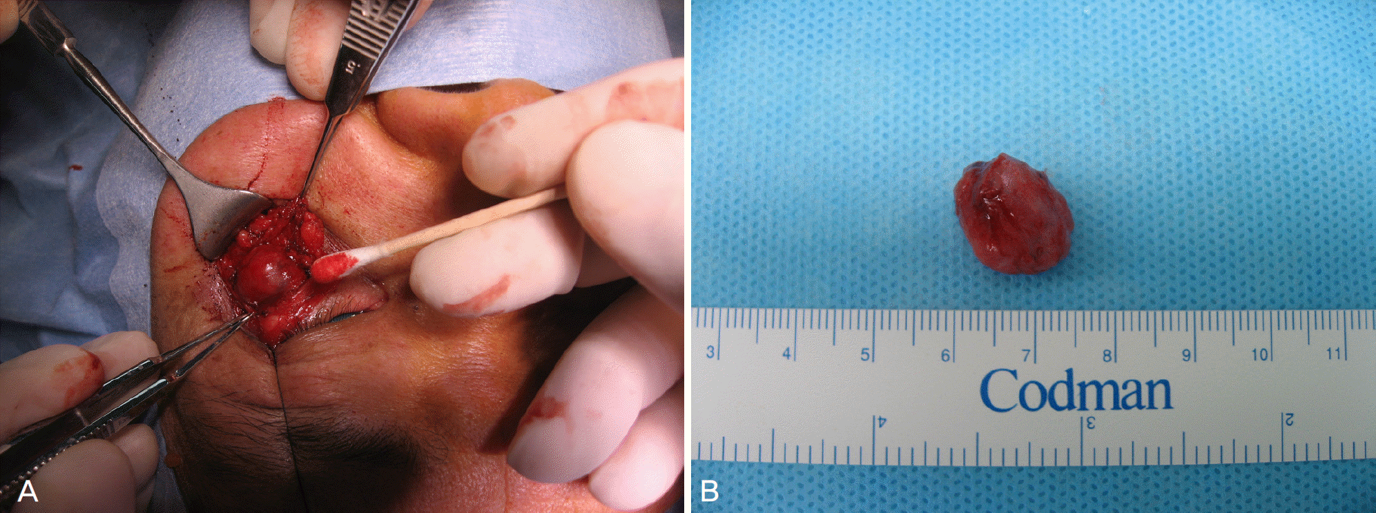
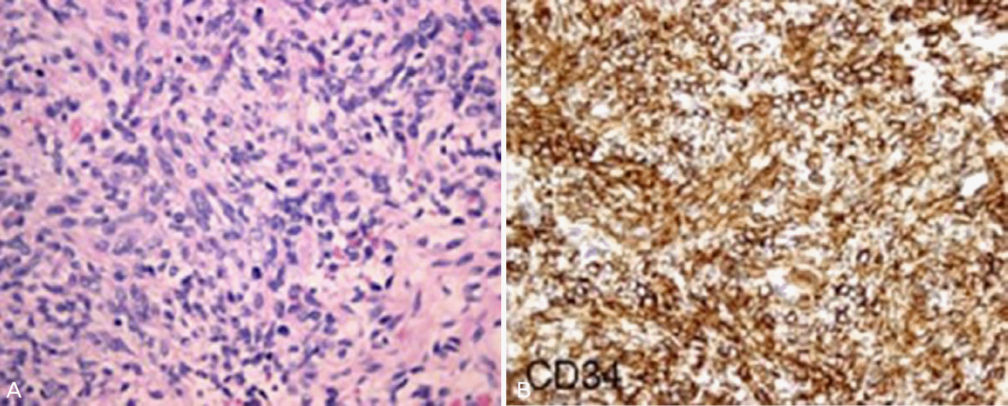
A 50-year-old man visited our clinic due to a slow progressing mass on the lateral side of the left eye for 1 year. The patient did not suffer from any discomfort or discharge from the mass. Slit lamp and other ocular examinations were unremarkable. Orbital MRI revealed a solid extra-conal enhanced mass that measured 13x11 mm adhering to the lateral wall of the left orbit. Total surgical excision was Performed under local anesthesia and tumor cells showed a strong and diffuse positivity for CD34 by immunohistochemistry. The findings were consistent with the diagnosis of orbital solitary tumor.
References
1. Bernardini FP, de Conciliis C, Schneider S, et al. Solitary fibrous tumor of the orbit. Is it rare? Report of a case series and review of the literature. Ophthalmology. 2003; 110:1442–8.
2. Leoncini G, Maio V, Puccioni M, et al. Orbital Solitary Fibrous tumor. A case report and review of the literature. Pathol Oncol Res. 2008; 14:213–7.

3. Schellini SA, Hoyama E, Marques ME, et al. Orbital Solitary Fibrous Tumor: Report of Two Cases and Literature Review. Jpn J Ophthalmol. 2003; 47:415–8.
4. Das JK, Sharma AS, Deka ACh, Das D. Solitary fibrous tumor of the orbit presenting in pregnancy. Indian J Ophthalmol. 2009; 57:238–40.
5. Pitchamuthu H, Gonzalez P, Kyle P, Roberts F. Fat-forming variant of solitary fibrous tumour of the orbit: the entity previously known as lipomatous haemangiopericytoma. Eye. 2009; 23:1479–81.
6. Lee NH, Ahn BC, Moon WS. A case of Solitary fibrous tumor of lacrimal gland. J Korean Ophthalmol. 2000; 41:2258–62.
7. Kang YK, Yoo HJ, Yum HK, Lee HS. Malignant solitary fibrous tumor of the pleura in mediastinum. Korean J Pathol. 1997; 31:351–6.
8. Westra WH, Gerald WL, Rosai J. Solitary fibrous tumor. Consistent CD34 immunoreactivity and occurrence in the orbit. Am J Surg Pathol. 1994; 18:992–8.
9. Lee JH, Sim SB, Park K, et al. Surgical therapy of solitary fibrous tumor of the parietal pleura. Korean Society for Thoracic and Cardiovascular Surgery. 1996; 29:798–801.
10. Dorfman DM, To K, Dickersin GR, et al. Solitary fibrous tumor of the orbit. Am J Surg Pathol. 1994; 18:281–7.
11. Cho NH, Kie JH, Yang WI, Jung WH. Solitary fibrous tumor with an unusual adenofibromatous feature in the lacrimal gland. Histopathology. 1998; 33:289–901.
Figure 1.
Preoperative external photographs of the fixed non-tender mass on the lateral side of the left lower eye-lid (black arrow) and lower lid entropion.
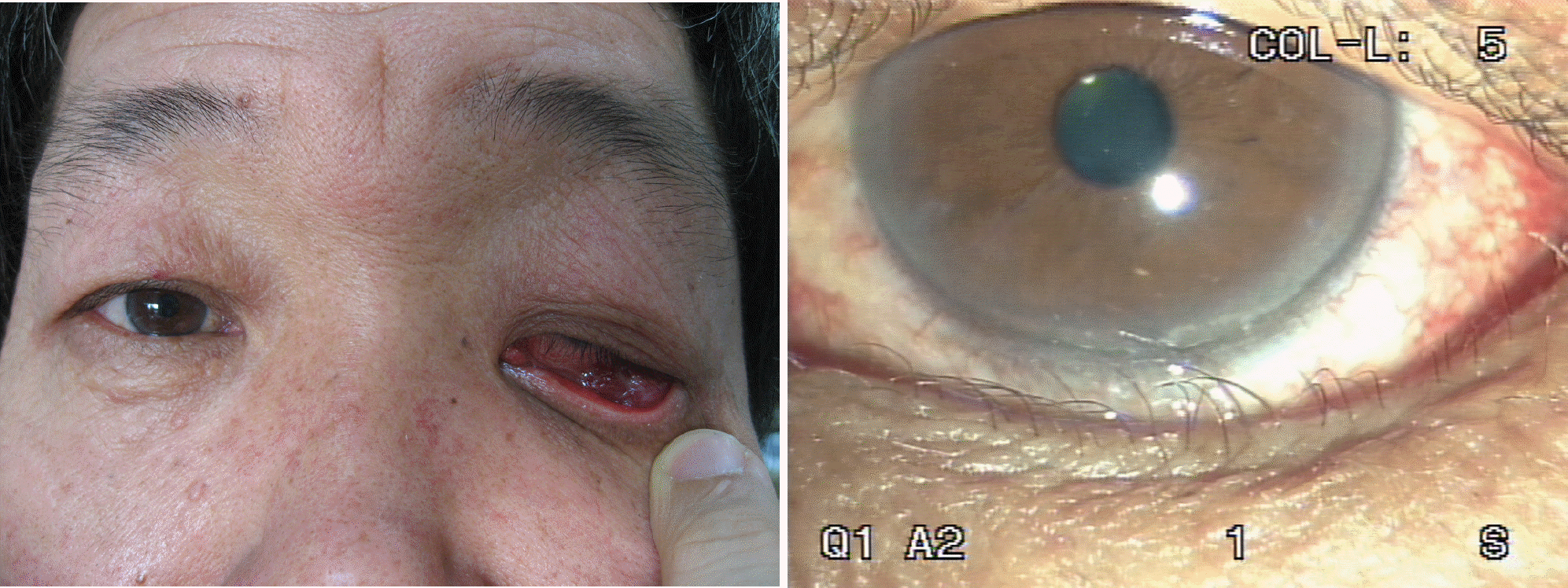
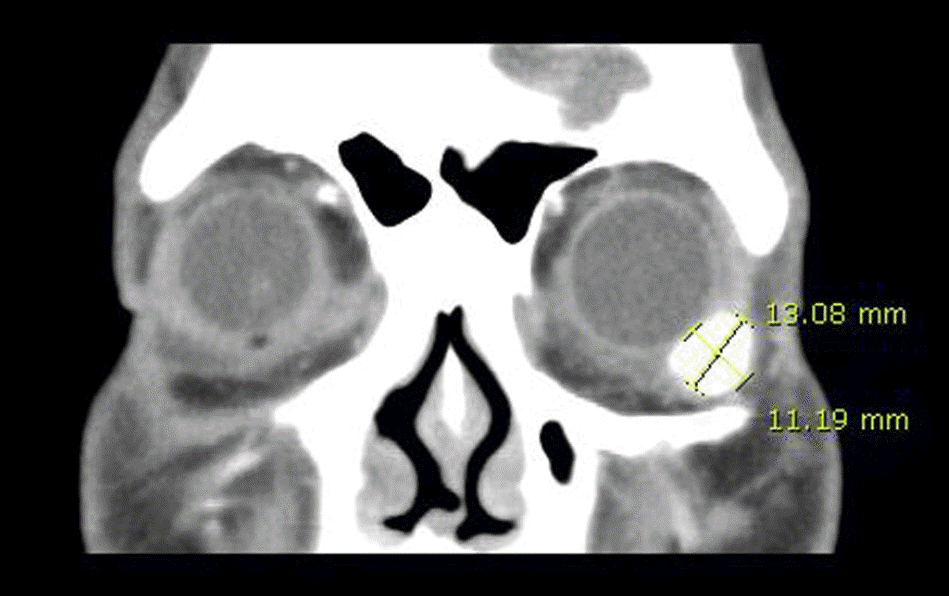
Figure 2.
Orbital CT (coronal view) finding shows a 13 mm×11 mm-sized well-circumscribed mass with ho-mogenous enhancement at the left lower orbital area.
 XML Download