

 PDF
PDF ePub
ePub Citation
 Print
Print


Abstract
Purpose
To report a case of subretinal hematoma secondary to polypoidal choroidal vasculopathy (PCV) misunderstood as a subretinal mass.
Case summary
A 73-year-old man with no specific medical history visited our clinic with decreased vision in the right eye. Slit-lamp examination revealed no specific findings for the anterior segment of the right eye. Upon fundus examination, an elevated macular lesion with some subretinal hemorrhages was observed, and a subretinal mass lesion was found on ultrasonography. After performing fluorescein angiography, indocyanine green angiography, and magnetic resonance imaging, we presumed that this lesion was a subretinal hematoma or ocular tumor and recommended observation. One month later, the subretinal mass had decreased in size. One year later, PCV with large retinal pigment epithelial detachment (RPED) was observed. After the intravitreal bevacizumab injection, RPED and macular edema were improved.
References
1. Yannuzzi LA, Sorenson J, Spaide RF, Lipson B. Idiopathic abdominal choroidal vasculopathy (IPCV). Retina. 1990; 10:1–8.
2. Yannuzzi LA, Wong DW, Sforzolini BS, et al. Polypoidal abdominal vasculopathy and neovascularized age-related macular degeneration. Arch Ophthalmol. 1999; 117:1503–10.
3. Yannuzzi LA, Ciardella A, Spaide RF, et al. The expanding abdominal spectrum of idiopathic polypoidal choroidal vasculopathy. Arch Ophthalmol. 1997; 115:478–85.
4. Spaide RF, Yannuzzi LA, Slakter JS, et al. Indocyanine green abdominal of idiopathic polypoidal choroidal vasculopathy. Retina. 1995; 15:100–10.
5. Moorthy RS, Lyon AT, Rabb MF, et al. Idiopathic polypoidal choroidal vasculopathy of the macula. Ophthalmology. 1998; 105:1380–5.

6. Scotto J, Fraumeni JF Jr, Lee JA. Melanomas of the eye and other noncutaneous sites: Epidemiologic aspects. J Natl Cancer Inst. 1976; 56:489–91.
7. Char DH, Stone RD, Irvine AR, et al. Diagnostic modalities in choroidal melanoma. Am J Ophthalmol. 1980; 89:223–30.
8. Scott IU, Murray TG, Hughes JR. Evaluation of imaging abdominal for detection of extraocular extension of choroidal melanoma. Arch Ophthalmol. 1998; 116:897–9.
9. Peyman GA, Mafee MF. Uveal melanoma and similar lesions: The role of magnetic resonance imaging and computed tomography. Radiol Clin North Am. 1987; 25:471–86.
10. Ahn KH, Kim KB, Joo CK. Two cases of malignant melanoma abdominal by MRI. J Korean Ophthalmol Soc. 1991; 32:825–31.
11. Tong JP, Chan WM, Liu DT, et al. Aqueous humor levels of abdominal endothelial growth factor and pigment epithelium derived factor in polypoidal choroidal vasculopathy and choroidal neovascularization. Am J Ophthalmol. 2006; 141:456–62.
12. Matsuoka M, Ogata N, Otsugi T, et al. Expression of pigment abdominal derived factor and vascular endothelial growth factor in choroidal neovascular membranes and polypoidal choroidal vasculopathy. Br J Ophthalmol. 2004; 88:809–15.
13. Lee MH, An JH, Lee JE, Oum BS. abdominal Efficacy of Intravitreal Bavacizumab for Polypoidal Choroidal Vasculopathy. J Korean Ophthalmol Soc. 2009; 50:51–60.
Figure 1.
A fundus photograph and images of the ultrasonography on the right eye. (A) A large sized (about 4×5 disc diameter) dark brown elevated subretinal mass lesion was found on the macula. (B) B-scan ultrasonography shows a well defined regular internal acoustic subretinal mass (red arrows) and A-scan ultrasonography shows middle amplitude internal reflectivity of the mass lesion.
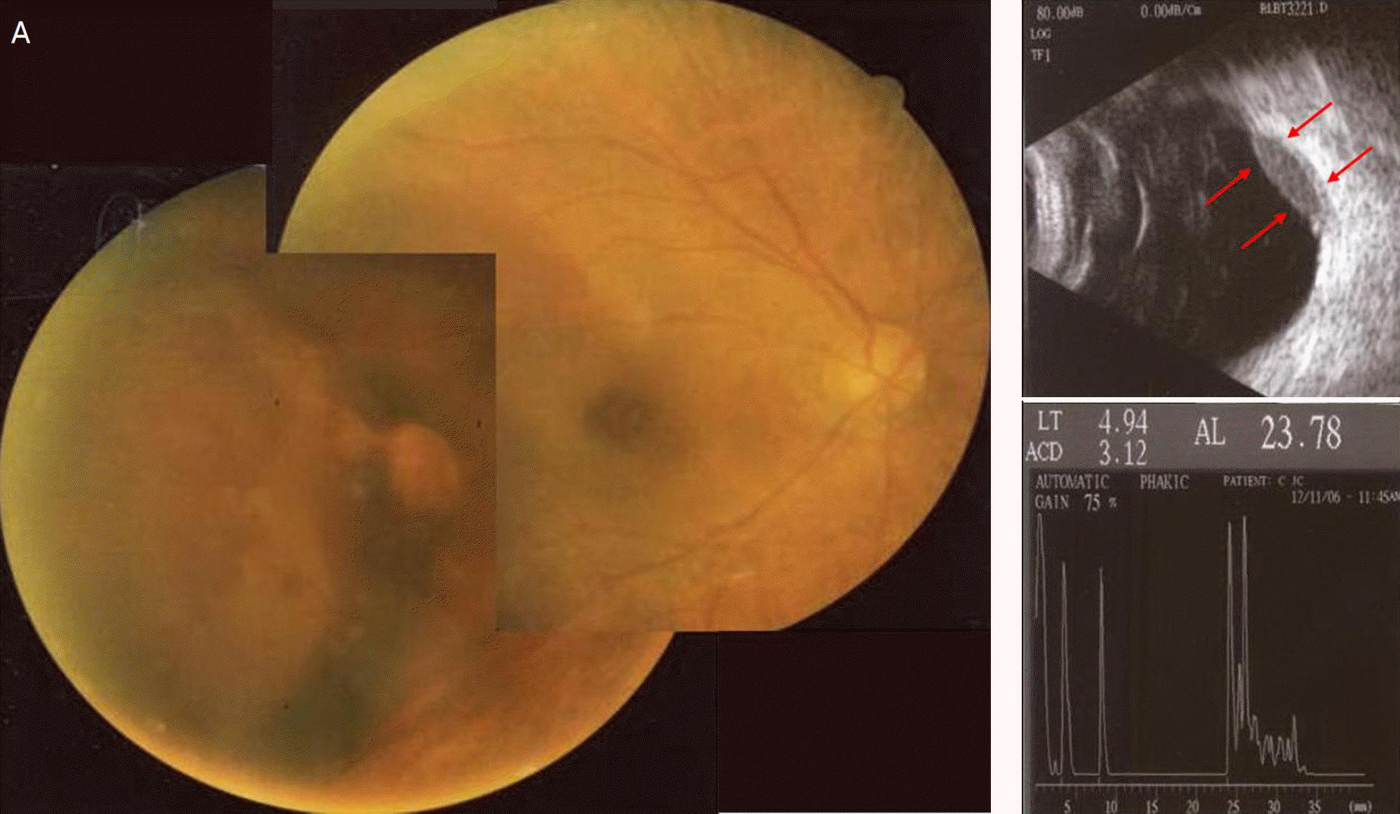
Figure 2.
Fluorescein angiogram and indocyanine green angiogram of the right eye. (A) Fluorescein angiogram shows large blocked choroidal fluorescence with multiple various sized hyperfluorescence. (B) Indocyanine green angiogram shows large blocked hypofluorescence with a small atypical hyperfluorescence in the temporal macula.
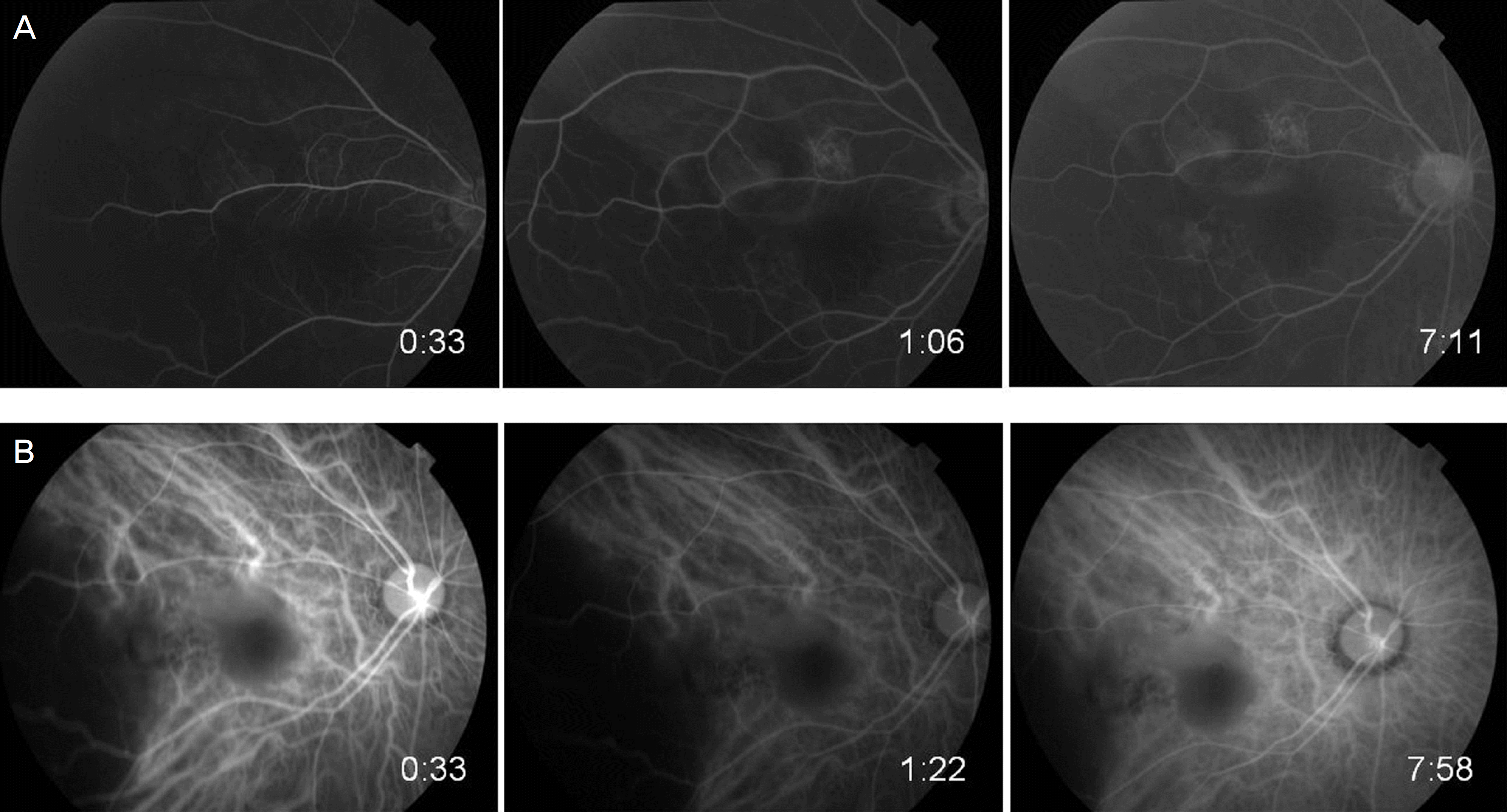
Figure 3.
Axial and coronal magnetic resonance images of the orbit. (A and B) T1-weighted images show increased signal intensity in the well defined crescent shaped mass (inside red loop) of the right eye. (C and D) T2-weighted images show decreased signal intensity in the mass (inside red loop) of the right eye.
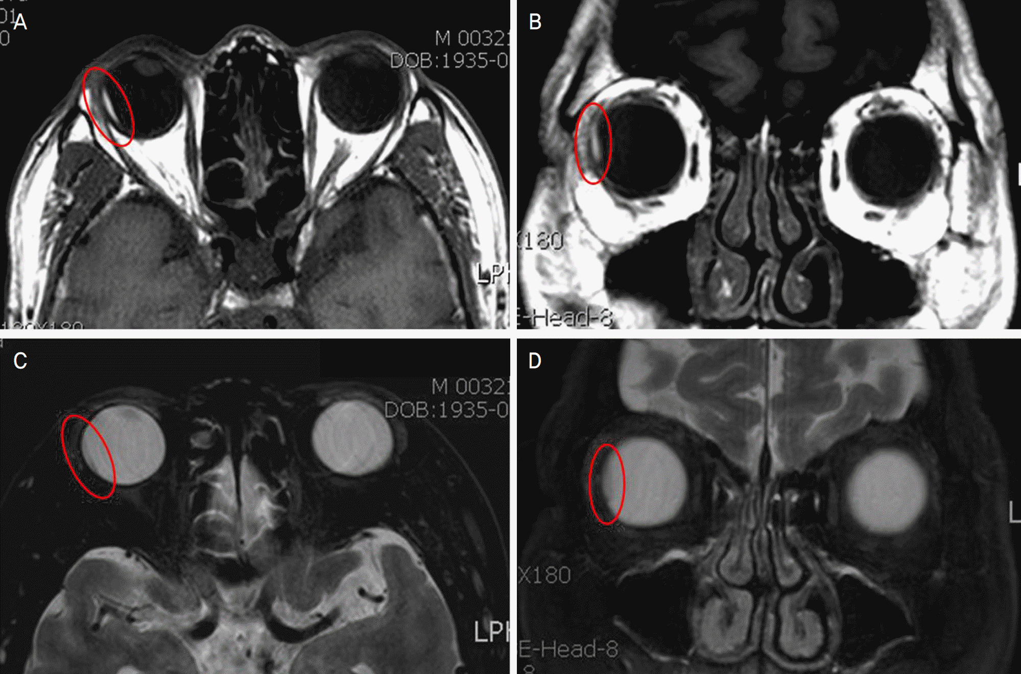
Figure 4.
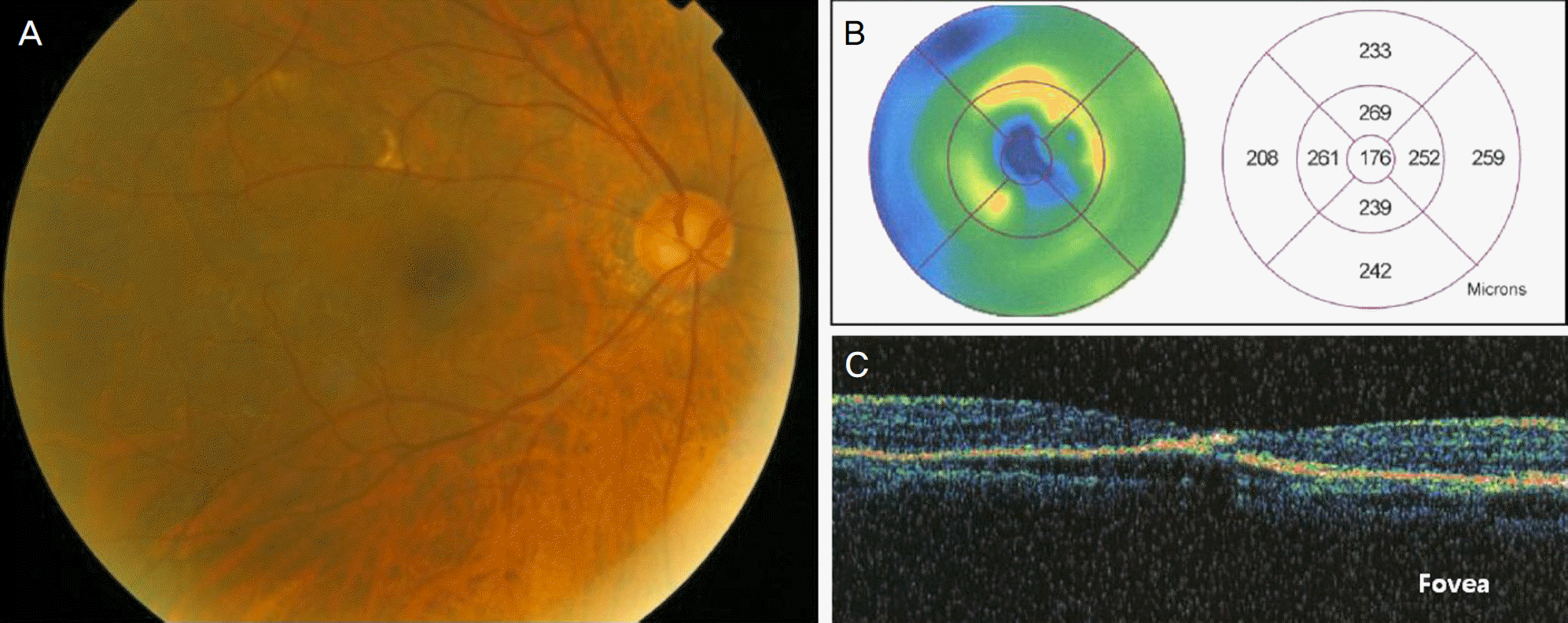
A fundus photograph (A) and an ultrasonographic image (B) on the right eye two months after the first visit. Subretinl hemorrhage was faint and subretinal mass lesion was decreased.
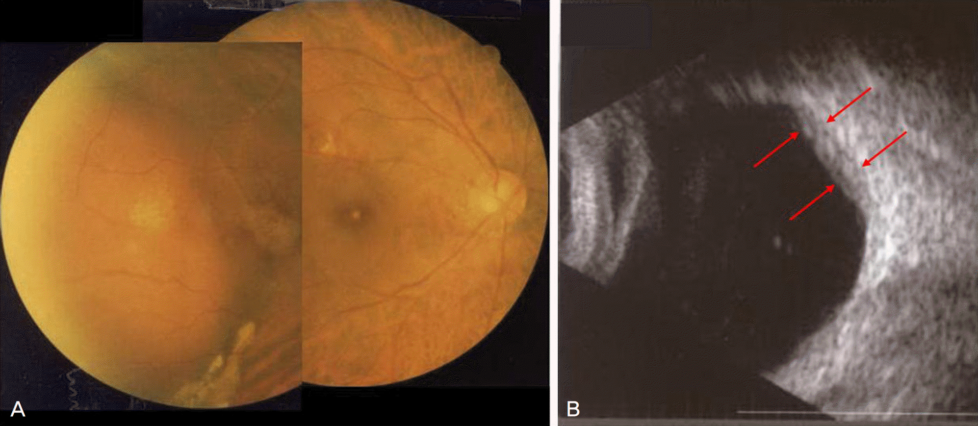
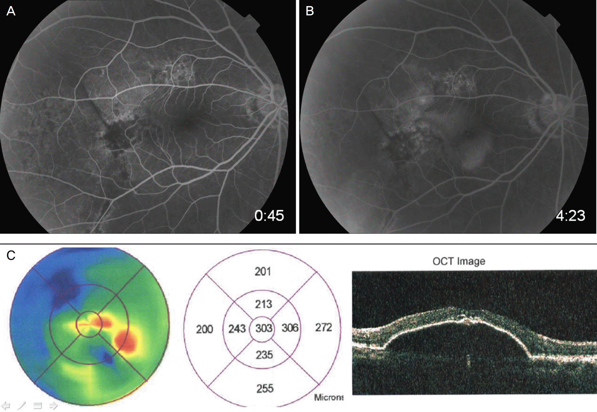
Figure 5.
Fluorescein angiogram and results of optical coherence tomography (OCT) on the right eye 1 year after the first visit. Early phase of fluorescein angiogram (A) shows multiple polypoidal hyperfluorescence and late phase of fluorescein angiogram (B) shows hyperfluorescent leakage and pooling on the posterior pole. Analysis and image of OCT scan (C) show macular edema and large retinal pigment epithelial detachment.
 XML Download