Abstract
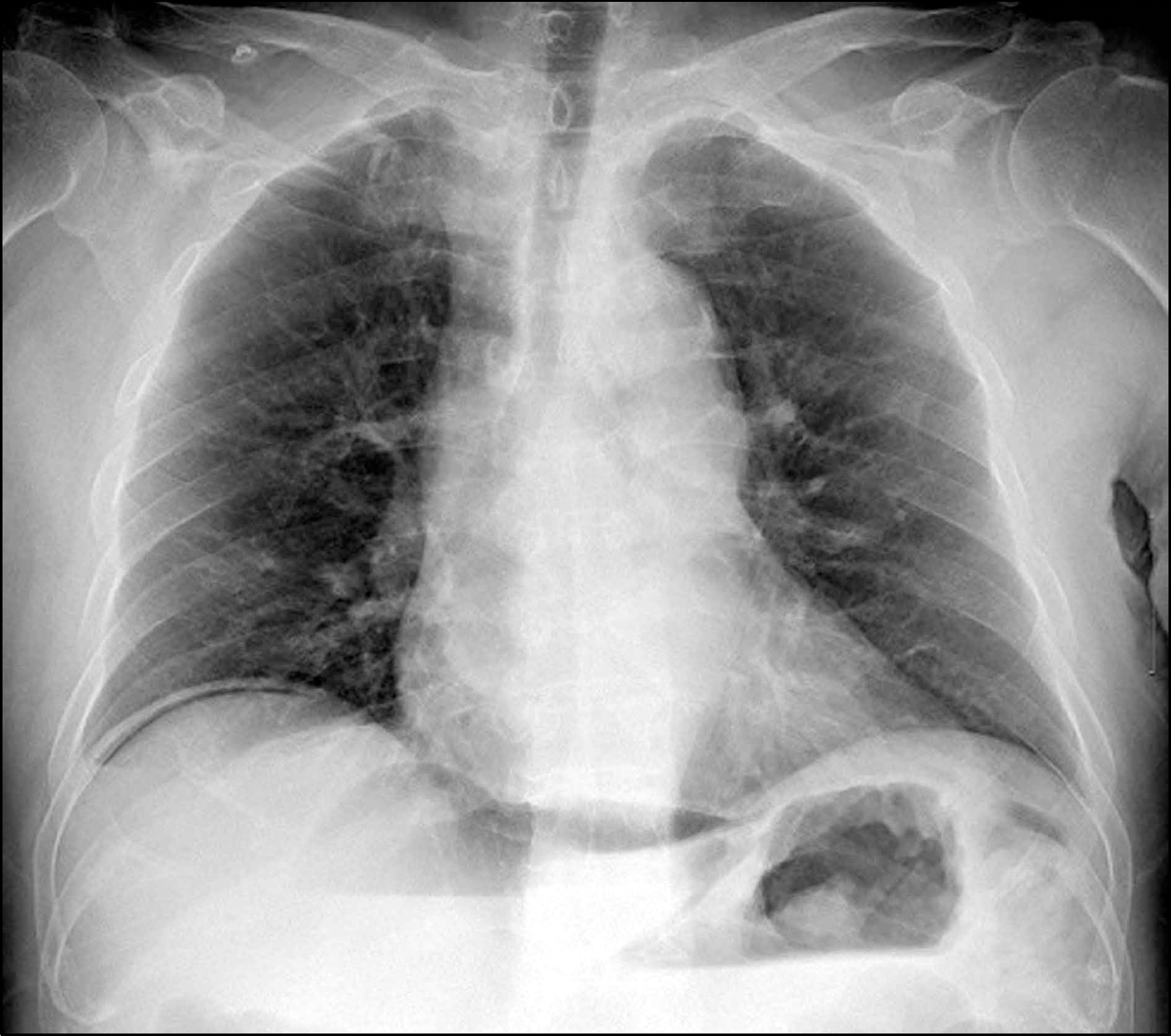
Spontaneous gastric perforation is a rare complication of gastric lymphoma that is potentially life threatening since it can progress to sepsis and multi-organ failure. Morbidity also increases due to prolonged hospitalization and delay in initiating chemotherapy. Therefore prompt diagnosis and appropriate treatment is critical to improve prognosis. A 64-year-old man presented to the emergency department with severe abdominal pain. Chest X-ray showed free air below the right diaphragm. Abdominal CT scan also demonstrated free air in the peritoneal cavity with large wall defect in the lesser curvature of gastric lower body. Therefore, the patient underwent emergency operation and primary closure was done. Pathologic specimen obtained during surgery was compatible to diffuse large B cell lymphoma. Fifteen days after primary closure, the patient received subtotal gastrectomy and chemotherapy was initiated after recovery. Patient is currently being followed-up at outpatient department without any particular complications. Herein, we report a rare case of gastric lymphoma that initially presented as peritonitis because of spontaneous gastric perforation.
References
1. Vaidya R, Habermann TM, Donohue JH, et al. Bowel perforation in intestinal lymphoma: incidence and clinical features. Ann Oncol. 2013; 24:2439–2443.

2. Gou HF, Zang J, Jiang M, Yang Y, Cao D, Chen XC. Clinical prognostic analysis of 116 patients with primary intestinal non-Hodgkin lymphoma. Med Oncol. 2012; 29:227–234.

3. Kim SG. Gastrointestinal lymphoma. Korean J Gastrointest Endosc. 2010; 41:1–4.
4. Aledavood A, Nasiri MR, Memar B, et al. Primary gastrointestinal lymphoma. J Res Med Sci. 2012; 17:487–490.
5. Shimada S, Gen T, Okamoto H. Malignant gastric lymphoma with spontaneous perforation. BMJ Case Rep. 2013. doi: 10.1136/bcr.05.2011.4251.

6. Nakamura S, Matsumoto T. Gastrointestinal lymphoma: recent advances in diagnosis and treatment. Digestion. 2013; 87:182–188.

7. Maisey N, Norman A, Prior Y, Cunningham D. Chemotherapy for primary gastric lymphoma: does inpatient observation prevent complications? Clin Oncol (R Coll Radiol). 2004; 16:48–52.

8. Ibrahim EM, Ezzat AA, El-Weshi AN, et al. Primary intestinal diffuse large B-cell non-Hodgkin's lymphoma: clinical features, management, and prognosis of 66 patients. Ann Oncol. 2001; 12:53–58.

9. Jhobta RS, Attri AK, Kaushik R, Sharma R, Jhobta A. Spectrum of perforation peritonitis in India–review of 504 consecutive cases. World J Emerg Surg. 2006; 1:26.
10. Ara C, Coban S, Kayaalp C, Yilmaz S, Kirimlioglu V. Spontaneous intestinal perforation due to non-Hodgkin's lymphoma: evaluation of eight cases. Dig Dis Sci. 2007; 52:1752–1756.

11. Papaxoinis G, Papageorgiou S, Rontogianni D, et al. Primary gastrointestinal non-Hodgkin's lymphoma: a clinicopathologic study of 128 cases in Greece. A Hellenic Cooperative Oncology Group study (HeCOG). Leuk Lymphoma. 2006; 47:2140–2146.

12. Daum S, Ullrich R, Heise W, et al. Intestinal non-Hodgkin's lymphoma: a multicenter prospective clinical study from the German Study Group on Intestinal non-Hodgkin's Lymphoma. J Clin Oncol. 2003; 21:2740–2746.

13. Hawkes EA, Wotherspoon A, Cunningham D. Diagnosis and management of rare gastrointestinal lymphomas. Leuk Lymphoma. 2012; 53:2341–2350.

14. Kitamura K, Yamaguchi T, Okamoto K, et al. Early gastric lymphoma: a clinicopathologic study of ten patients, literature review, and comparison with early gastric adenocarcinoma. Cancer. 1996; 77:850–857.

15. Caletti G, Fusaroli P, Togliani T, Bocus P, Roda E. Endosonography in gastric lymphoma and large gastric folds. Eur J Ultrasound. 2000; 11:31–40.

16. Wang HW, Yang W, Wang L, Lu YL, Lu JY. Composite diffuse large B-cell lymphoma and classical Hodgkin's lymphoma of the stomach: case report and literature review. World J Gastroenterol. 2013; 19:6304–6309.

17. Avilés A, Nambo MJ, Neri N, et al. The role of surgery in primary gastric lymphoma: results of a controlled clinical trial. Ann Surg. 2004; 240:44–50.
18. Binn M, Ruskoné-Fourmestraux A, Lepage E, et al. Surgical re-section plus chemotherapy versus chemotherapy alone: comparison of two strategies to treat diffuse large B-cell gastric lymphoma. Ann Oncol. 2003; 14:1751–1757.
Fig. 2.
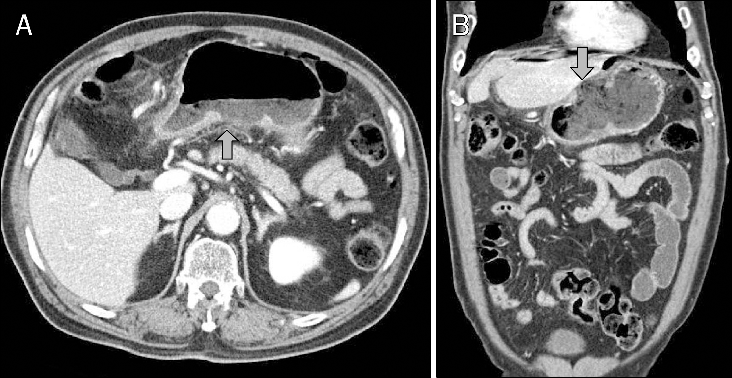
Abdominal CT scan findings. A large wall defect (arrows) and edema-tous thickening are noted in the lesser curvature of gastric lower body (A, transverse view; B, coronal view).

Fig. 3.
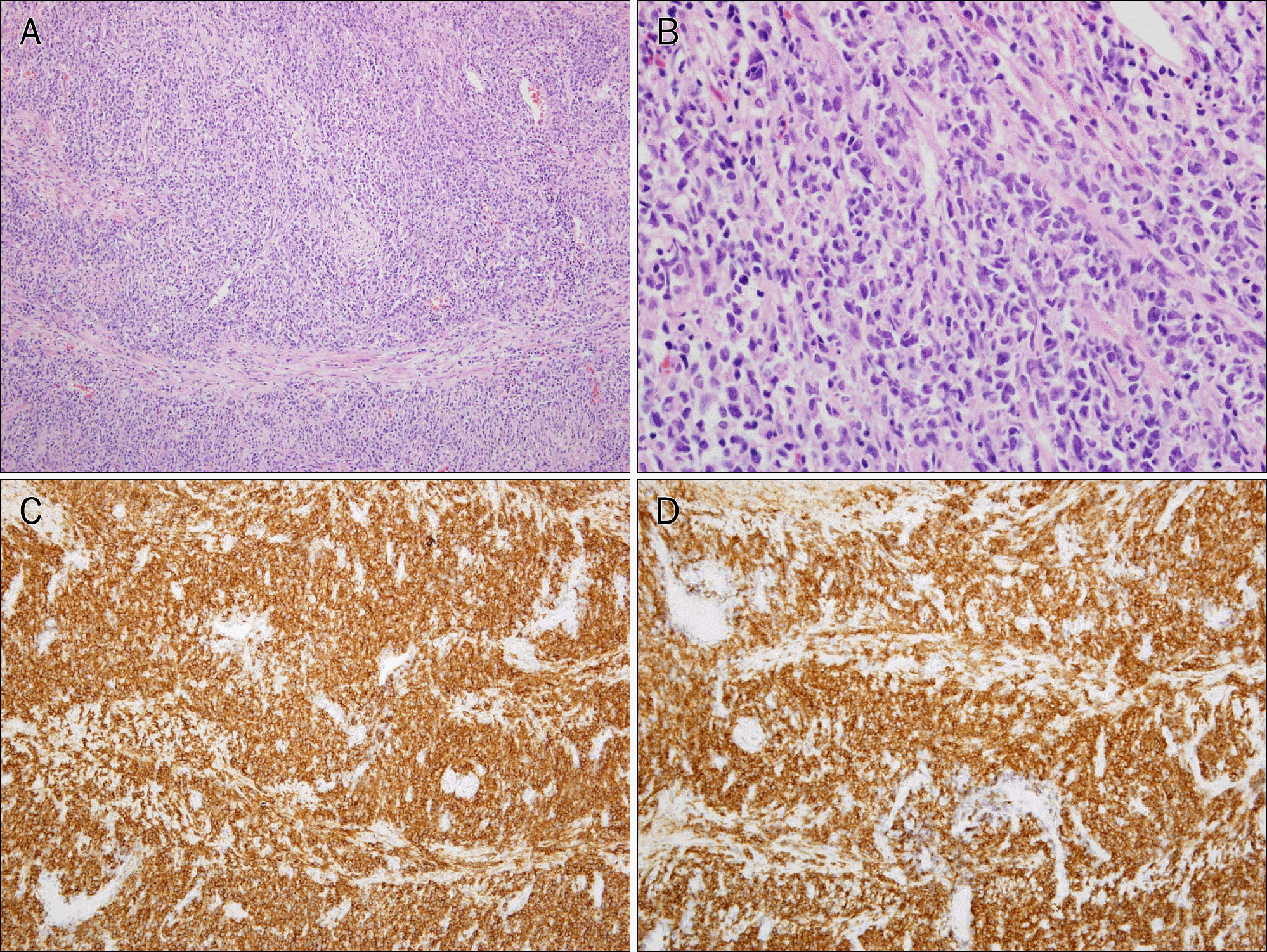
Microscopic findings. Diffuse proliferation of medium to large sized lymphocytes having vesicular nuclei with smooth chromatin and scant cytoplasm are observed (H&E; A, ×100; B, ×400). Immunohistochemistry staining shows that the tumor cells are positive for CD 20 (C, ×100) and leukocyte common antigen (D, ×100).

Fig. 4.
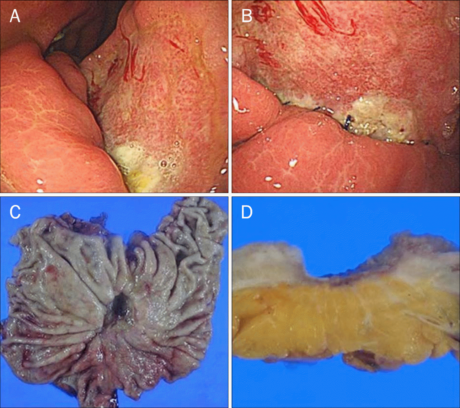
Endoscopic and gross findings.(A) An ulcerative lesion extending from the lesser curvature of gastric lower body to the gastric angle can be seen.(B) Suture line is observed on the anterior side of the lesion. (C, D) The resected specimen demonstrates a ulceroinfiltrative lesion containing 3.0×1.8 cm sized tumor in the lesser curvature of lower body.


 PDF
PDF ePub
ePub Citation
 Print
Print


 XML Download