

 PDF
PDF ePub
ePub Citation
 Print
Print


Abstract
Hepatocellular carcinoma (HCC) is a critical global health issue and the third most common cause of cancer-related deaths worldwide. The majority of patients who present HCC are already at an advanced stage and their tumors are unresectable. Sorafenib is a multikinase inhibitor of the vascular endothelial growth factor pathway and was recently introduced as a therapy for advanced HCC. Furthermore, studies have shown that oral sorafenib has beneficial effects on survival. However, many patients experience diverse side effects, and some of these are severe. Liver abscess development has not been previously documented to be associated with sorafenib administration in HCC. Here, we report the case of a HCC patient that developed a liver abscess while being treated with sorafenib.
References
1. Miura H, Miyazaki T, Kuroda M, et al. Increased expression of vascular endothelial growth factor in human hepatocellular carcinoma. J Hepatol. 1997; 27:854–861.

2. Torimura T, Sata M, Ueno T, et al. Increased expression of vascular endothelial growth factor is associated with tumor progression in hepatocellular carcinoma. Hum Pathol. 1998; 29:986–991.
3. Wilhelm SM, Carter C, Tang L, et al. BAY 43–9006 exhibits broad spectrum oral antitumor activity and targets the RAF/MEK/ERK pathway and receptor tyrosine kinases involved in tumor progression and angiogenesis. Cancer Res. 2004; 64:7099–7109.
4. Llovet JM, Ricci S, Mazzaferro V, et al. SHARP Investigators Study Group. Sorafenib in advanced hepatocellular carcinoma. N Engl J Med. 2008; 359:378–390.
5. Cheng AL, Kang YK, Chen Z, et al. Efficacy and safety of sorafenib in patients in the Asia-Pacific region with advanced hepatocellular carcinoma: a phase III randomised, double-blind, placebo-controlled trial. Lancet Oncol. 2009; 10:25–34.
6. Rahimian J, Wilson T, Oram V, Holzman RS. Pyogenic liver abscess: recent trends in etiology and mortality. Clin Infect Dis. 2004; 39:1654–1659.
7. Brook I, Frazier EH. Microbiology of liver and spleen abscesses. J Med Microbiol. 1998; 47:1075–1080.
8. Lieuw-a-Fa M, Peringa J, Leeksma O, Terpstra W. Sepsis from liver abscesses in metastatic colorectal carcinoma after chemo-immunotherapy. J Clin Oncol. 2008; 26:1381–1382.
9. Chen C, Chen PJ, Yang PM, et al. Clinical and microbiological features of liver abscess after transarterial embolization for hepatocellular carcinoma. Am J Gastroenterol. 1997; 92:2257–2259.
10. Ong GY, Changchien CS, Lee CM, et al. Liver abscess complicating transcatheter arterial embolization: a rare but serious complication. A retrospective study after 3878 procedures. Eur J Gastroenterol Hepatol. 2004; 16:737–742.
11. Kim MH, Choi MS, Choi YS, et al. Clinical features of liver abscess developed after radiofrequency ablation and transarterial chemoembolization for hepatocellular carcinoma. Korean J Hepatol. 2006; 12:55–64.
12. Rhim H, Yoon KH, Lee JM, et al. Major complications after radiofrequency thermal ablation of hepatic tumors: spectrum of imaging findings. Radiographics. 2003; 23:123–134. discussion 134–136.
13. Livraghi T, Solbiati L, Meloni MF, Gazelle GS, Halpern EF, Goldberg SN. Treatment of focal liver tumors with percutaneous radiofrequency ablation: complications encountered in a multicenter study. Radiology. 2003; 226:441–451.
14. Kobayashi S, Nakanuma Y, Terada T, Matsui O. Postmortem survey of bile duct necrosis and biloma in hepatocellular carcinoma after transcatheter arterial chemoembolization therapy: relevance to microvascular damages of peribiliary capillary plexus. Am J Gastroenterol. 1993; 88:1410–1415.
15. Kim YK, Yu SE, Hong CK, et al. Liver abscess formation in non-tumorous parenchyma after transcatheter arterial chemoembolization (TACE) for the treatment of hepatocellular carcinoma associated with pneumobilia. Korean J Hepatol. 2001; 7:189–194.
16. Everson TC, Cole WH. Spontaneous regression of cancer. Philadelphia: WB Saunders;1966. p. 6–7.
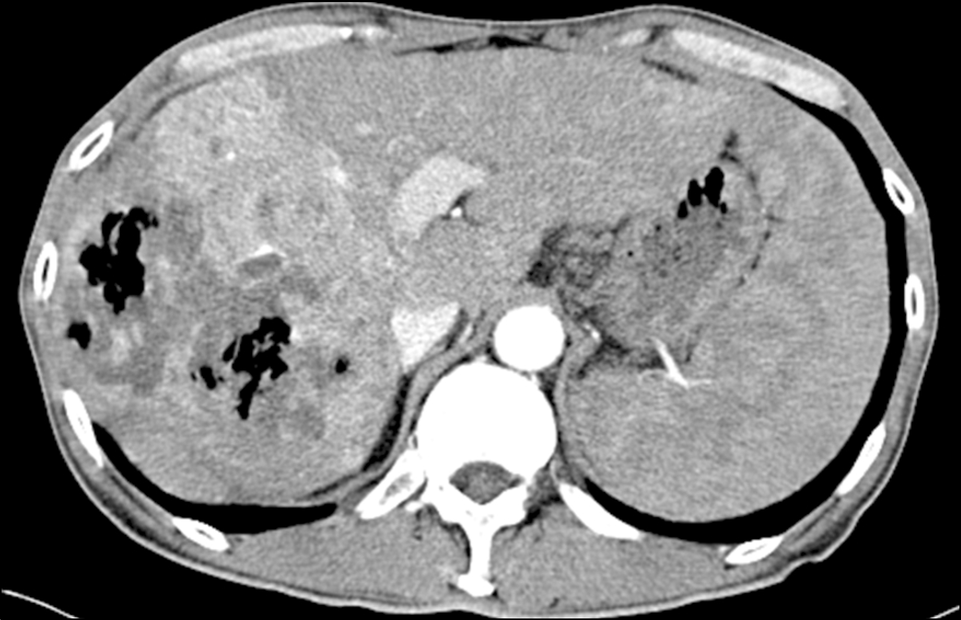
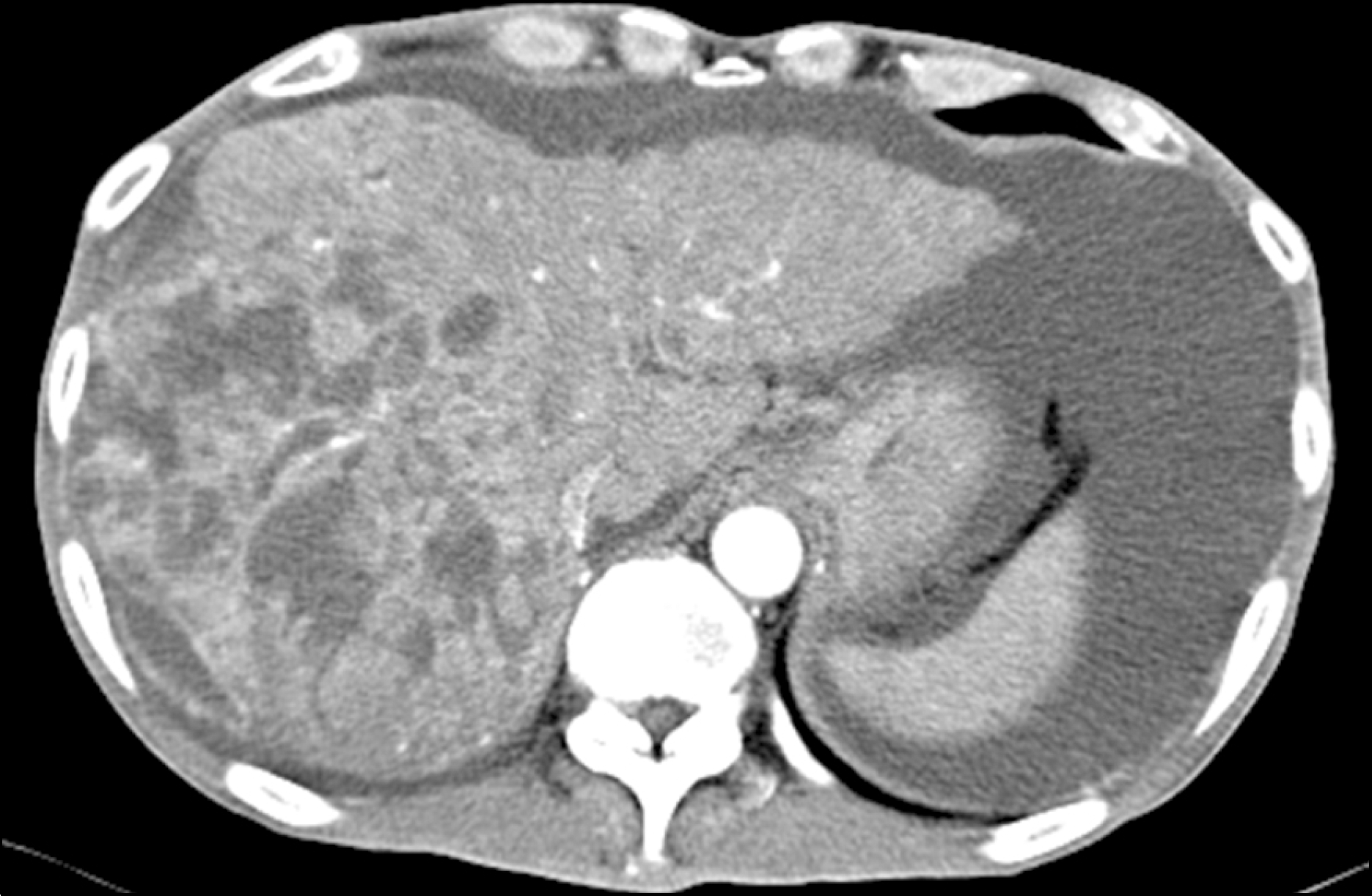
Fig. 1.
Liver CT scan at the time of hepatocellular carcinoma diagnosis, showing a 10-cm sized high density mass in the arterial phase (arrow) and washout in the portal venous phase, with portal vein thrombosis (arrow head) in the right lobe of the liver. (A) Arterial phase. (B) Portal venous phase.

 XML Download