REFERENCES
1. Garnick M, Lokich JJ. Primary malignant melanoma of the rectum: rationale for conservative surgical management. J Surg Oncol. 1978; 10:529–531.

2. Slingluff CL Jr, Vollmer RT, Seigler HF. Anorectal melanoma: clinical characteristics and results of surgical management in twenty-four patients. Surgery. 1990; 107:1–9.
3. Ojima Y, Nakatsuka H, Haneji H, et al. Primary anorectal malignant melanoma: report of a case. Surg Today. 1999; 29:170–173.

4. Husa A, Hö ckerstedt K. Anorectal malignant melanoma. A report of fourteen cases. Acta Chir Scand. 1974; 140:68–72.
6. Pantalone D, Taruffi F, Paolucci R, Liguori P, Rastrelli M, Andreoli F. Malignant melanoma of the rectum. Eur J Surg. 2000; 166:583–584.
7. Cooper PH, Mills SE, Allen MS Jr. Malignant melanoma of the anus: report of 12 patients and analysis of 255 additional cases. Dis Colon Rectum. 1982; 25:693–703.
8. Tanaka S, Ohta T, Fugimoto T, Makino Y, Murakami I. Endoscopic mucosal resection of primary anorectal malignant melanoma: a case report. Act Med Okayama. 2008; 62:421–424.
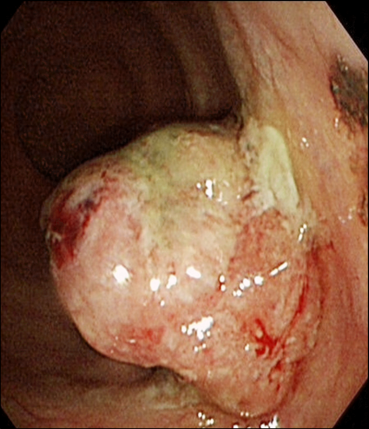
Fig. 1.
Endoscopic finding of the polypoid mass. There was about 3 cm sized fungating mass at the rectum. At the top of the mass, slightly black colored base was noted.

Fig. 2.
Preoperative radiologic findings. (A) At the rectum, about 3 cm sized fungating mass was noted. (B) There was one hepatic cyst, but no metastasis.


 PDF
PDF ePub
ePub Citation
 Print
Print


 XML Download