



 PDF
 ePub
 Citation
 Print


Abstract
Nephrogenic diabetes insipidus (DI) is characterized by insensitivity of the distal nephron to vasopressin (ADH) and the inability to concentrate urine, which leads to excreting excessive quantities of urine. Upper tract dilation secondary to polyuria was previously shown to be associated with nephrogenic DI. We report here on 3 males with nephrogenic DI that caused voiding difficulty as well as massive nonobstructive dilation of the urinary tract.
REFERENCES
1. Shalev H, Romanovsky I, Knoers NV, Lupa S, Landau D. Bladder function impairment in aquaporin-2 defective nephrogenic diabetes insipidus. Nephrol Dial Transplant. 2004; 19:608–13.

2. Tank ES, Alexander SR, Craven RM. Polyuric megalocystis. J Urol. 1980; 124:692–4.
3. Streitz JM Jr, Sreitz JM. Polyuric urinary tract dilatation with renal damage. J Urol. 1988; 139:784–5.
4. Boyd SD, Raz S, Ehrlich RM. Diabetes insipidus and nonobstructive dilation of urinary tract. Urology. 1980; 16:266–9.
5. McGuire EJ, Woodside JR, Borden TA, Weiss RM. Prognostic value of urodynamic testing in myelodysplastic patients. J Urol. 1981; 126:205–9.
6. Park CY, Kim HS. Treatment of renal injury in a patient presenting pituitary diabetes insipidus associated with bilateral hydronephrosis: a case report. Korean J Urol. 1994; 35:74–81.
7. Kim CI, Lee BS, Suh JK, Lee MS. A case of nephrogenic diabetes insipidus associated with hydronephrosis. Korean J Urol. 1983; 24:1101–5.
8. Zender HO, Ruedin P, Moser F, Bolle JF, Leski M. Traumatic rupture of the urinary tract in a patient presenting nephrogenic diabetes insipidus associated with hydronephrosis and chronic renal failure: case report and review of the literature. Clin Nephrol. 1992; 38:196–202.
Fig. 1.
Voiding cystourethrogram (A). The “christmas tree” appearance of the bladder was seen on ultrasonography (B). Severe hydronephrosis with dilation of the bladder and diatal ureter was seen.
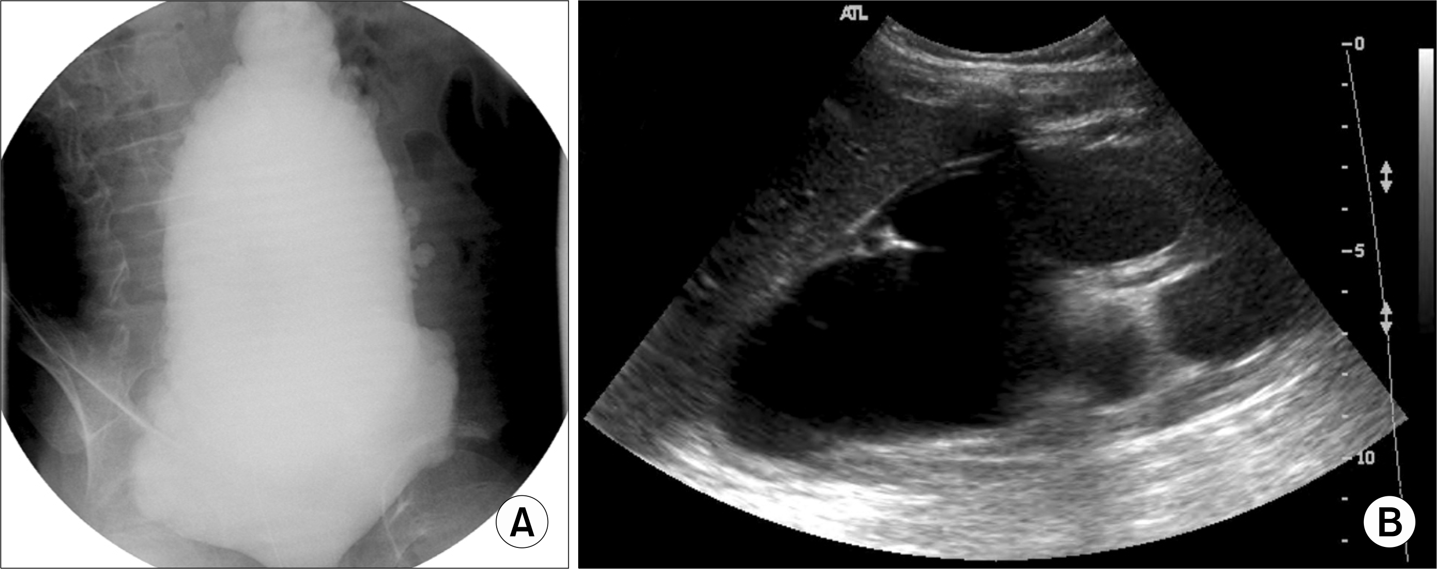
Fig. 2.
(A) The uroflowmetry study revealed that the patient was voiding by abdominal straining. (B) The filling cystometrogram (CMG) shows a large bladder capacity with decreased sensation. (C) The voiding CMG demonstrates slightly weak detrusor contractility with abdominal straining.
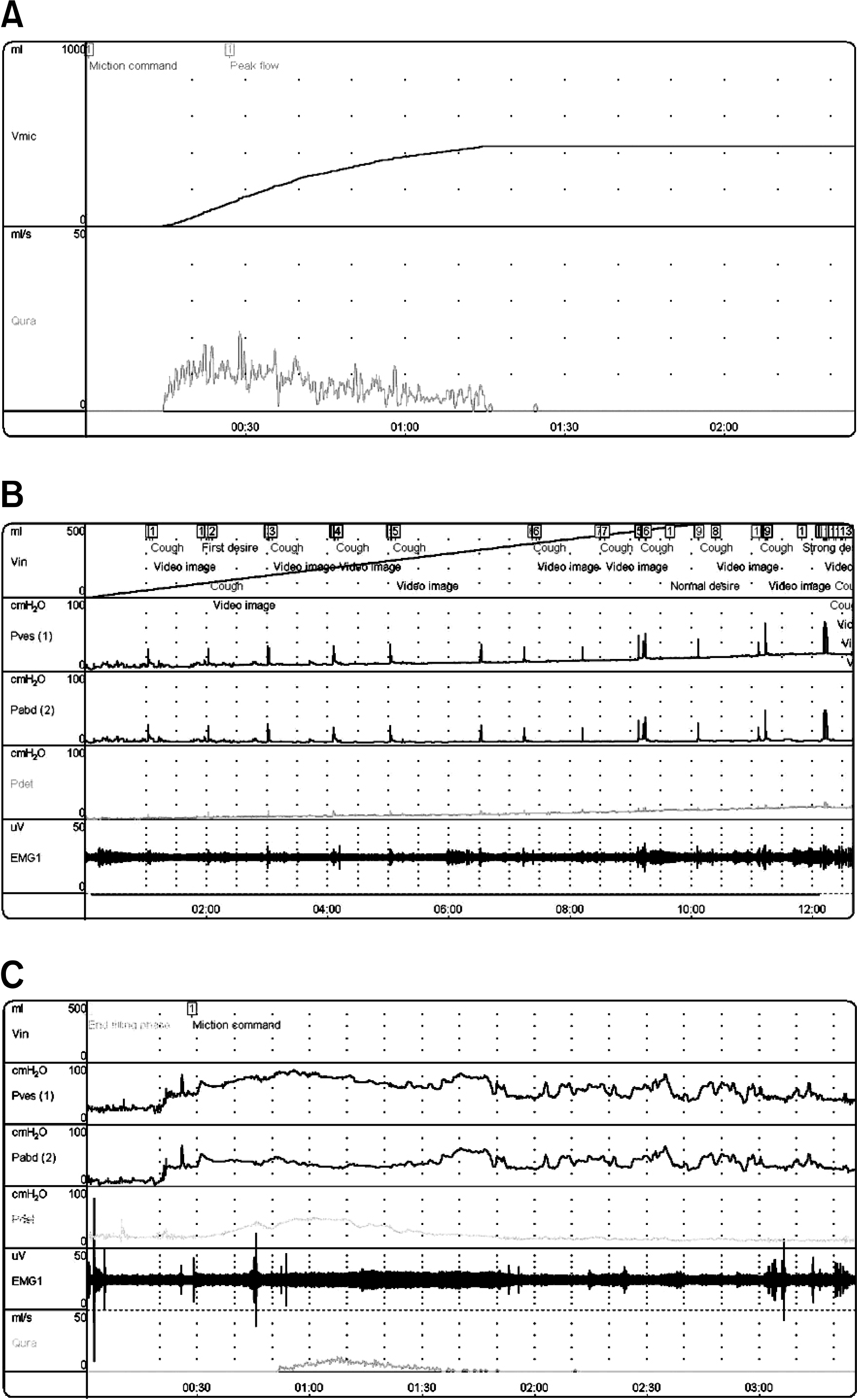
Table 1.
Fluoroscopic urodynamic findings and the upper tract imaging study
 XML Download