

 PDF
PDF ePub
ePub Citation
 Print
Print


Abstract
Urethral diverticulum is a relatively uncommon abnormality for a man. We report here on a case of acquired anterior urethral diverticulum with giant calculi and cutaneous fistula in a 64-years old male patient who had suffered from urethritis 10 years previously. Retrograde urethrography and urethroscopy confirmed the anterior urethral diverticulum with giant calculi, and these procedures ruled out the presence of any associated obstructive urethral anomalies. We performed urethral diverticulectomy, with removal of the calculi, and then we performed urethroplasty.
References
1. Marya S, Kumar S, Singh S. Acquired male urethral diverticulum. J Urol. 1977; 118:765–6.

2. Mohan V, Gupta SK, Cherian J, Tripathi VN, Sharma BB. Urethral diverticulum in male subjects: report of 5 cases. J Urol. 1980; 123:592–4.
3. Ortlip S, Gonzalez R, Williams RD. Diverticula of the male urethra. J Urol. 1980; 124:350–5.
4. Mandler J, Pool T. Primary diverticulum of the male urethra. J Urol. 1966; 96:336–8.
5. Lee SI, Jeong TY, Shim HY. Male urethral diverticulum combined with stone and urethrocutaneou fistula. Korean J Urol. 2002; 43:350–2.
6. Ahn DW, Kim KS, Oh MM, Yang KY. A case of male urethral diverticulum with giant calculi. Korean J Urol. 1999; 40:1723–5.
7. Lee YH, Park CB, An SS, Jang YI, Oh KJ, Kim KH. A case of urethral diverticulum combined with giant stone. Korean J Urol. 1995; 36:1165–7.
8. Kim KN, Cha YI, Lee KP. A case of urethral diverticulum combined with stone. Korean J Urol. 1970; 11:255–7.
9. Ginesin Y, Bolkier M, Nachmias BJ, Levin DR. Primary giant calculus in urethral diverticulum. Urol Int. 1988; 43:47–8.
10. Shinha A, Rintoul RF. Giant calculus in urethral diverticulum. Br J Urol. 1982; 54:62.
Fig. 1.
A large stony hard mass was located at the ventral portion of the penile shaft (A). Note the fistulous orifice at the mass lesion of the penile shaft (B).
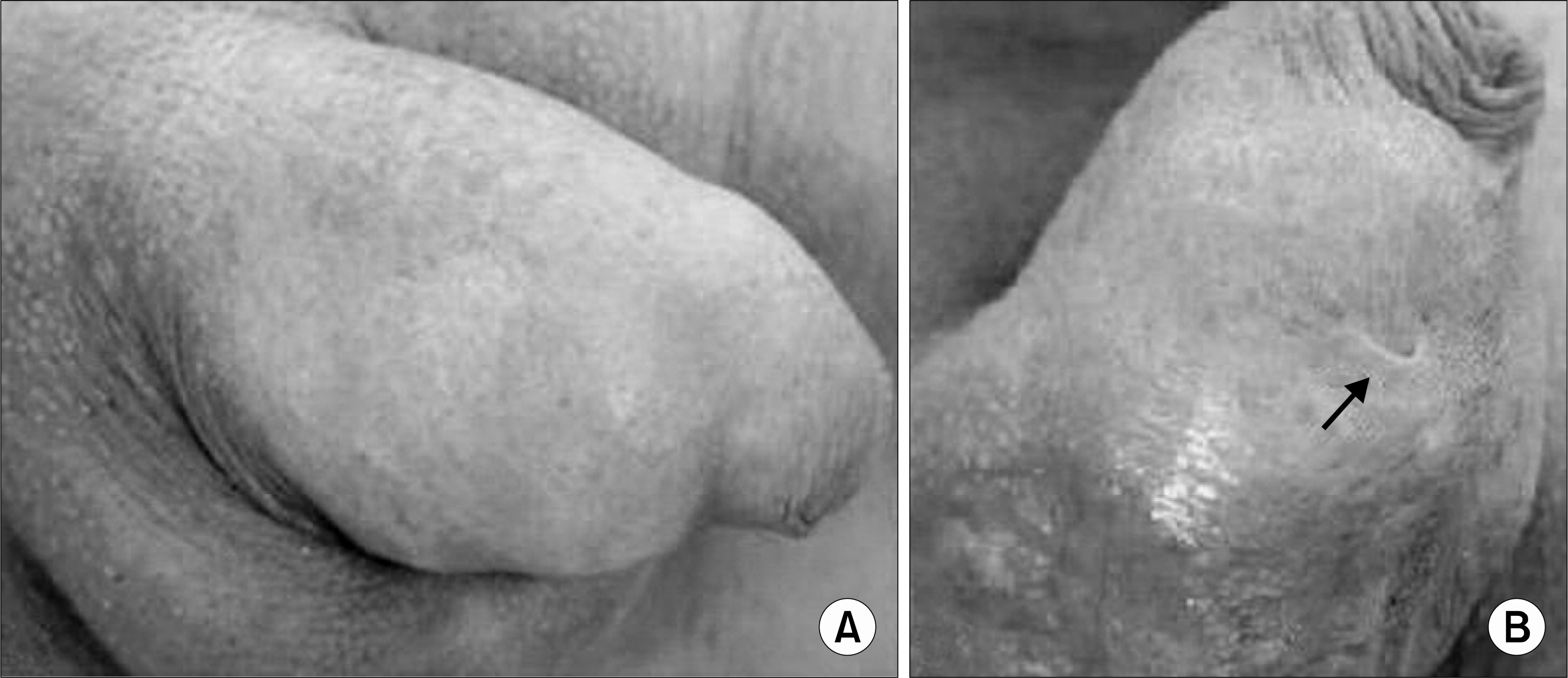
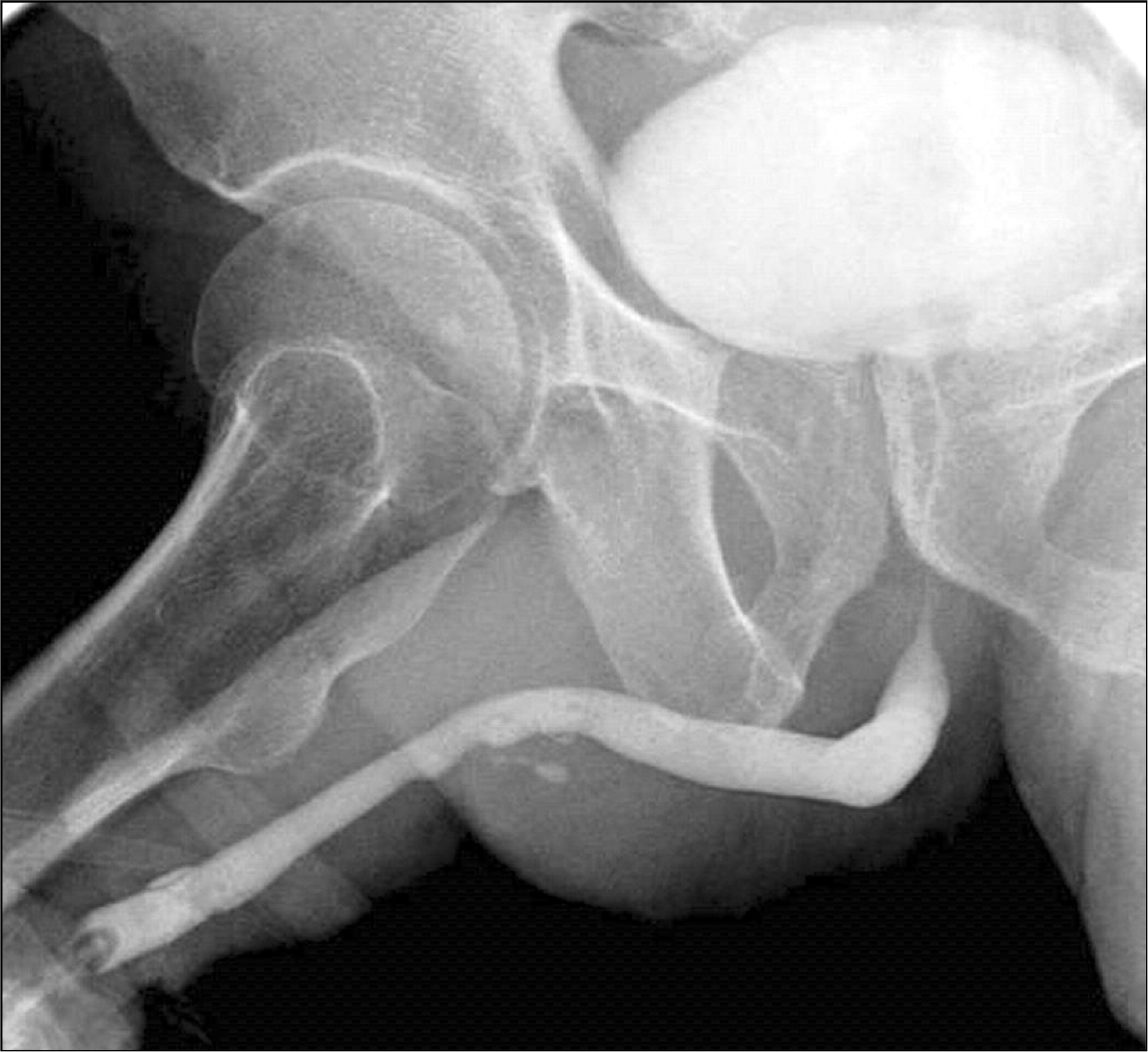
Fig. 2.
Retrograde urethrography shows the urethral diverticulum with giant stone and cutaneous fistula. Note the leakage of contrast media through the cutaneous fistula (A). Urethroscopy shows the multiple urethral diverticular ostia at the mid portion of the anterior urethra (B).


 XML Download