REFERENCES
1). Ghanem I. The management of osteoid osteoma: updates and controversies. Curr Opin Pediatr. 2006. 18:36–41.

2). Gaeta M., Minutoli F., Pandolfo I., Vinci S., D'Andrea L., Blandino A. Magnetic resonance imaging findings of osteoid osteoma of the proximal femur. Eur Radiol. 2004. 14:1582–9.

3). von Chamier G., Holl-Wieden A., Stenzel M., Raab P., Darge K., Girschick HJ, et al. Pitfalls in diagnostics of hip pain: osteoid osteoma and osteoblastoma. Rheumatol Int. 2010. 30:395–400.

4). Alani WO., Bartal E. Osteoid osteoma of the femoral neck simulating and inflammatory synovitis. Clin Orthop Relat Res. 1987. 223:308–12.
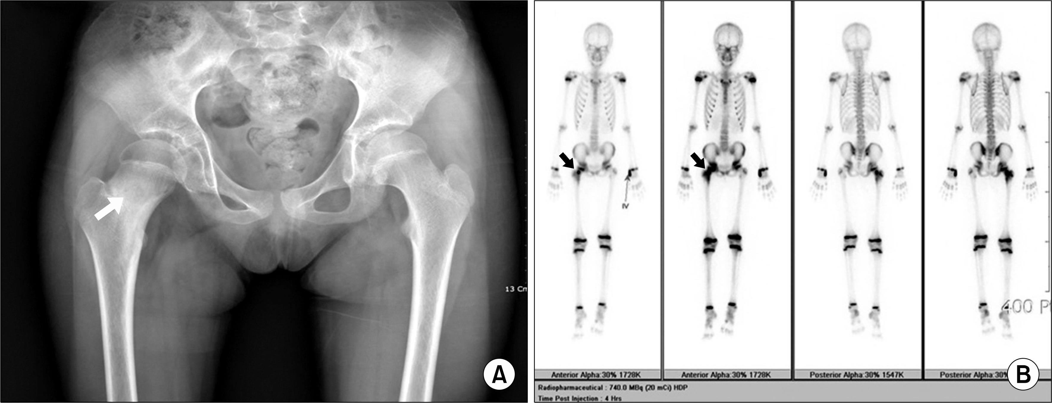
Fig. 1.
(A) Hip X-ray shows small radiolucent lesion with surrounding sclerosis (white arrow), (B) Whole body bone scan shows increased radioisotope uptake in the right femoral neck with reactive bone change (black arrow).


 PDF
PDF ePub
ePub Citation
 Print
Print


 XML Download